. 2014:2014:584314.
doi: 10.1155/2014/584314. Epub 2014 Sep 3.

Abeta(1-42) enhances neuronal excitability in the CA1 via NR2B subunit-containing NMDA receptors

Affiliations

Abeta(1-42) enhances neuronal excitability in the CA1 via NR2B subunit-containing NMDA receptors

Edina Varga et al. Neural Plast. 2014.

Abstract

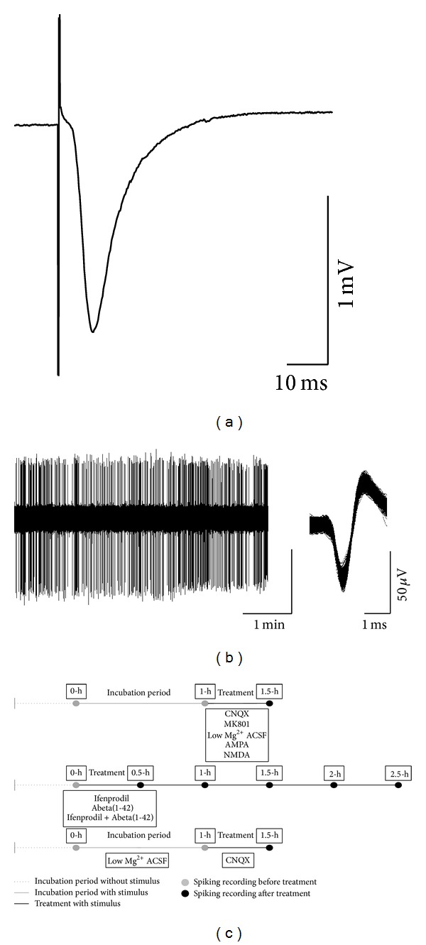
Neuronal hyperexcitability is a phenomenon associated with early Alzheimer's disease. The underlying mechanism is considered to involve excessive activation of glutamate receptors; however, the exact molecular pathway remains to be determined. Extracellular recording from the CA1 of hippocampal slices is a long-standing standard for a range of studies both in basic research and in neuropharmacology. Evoked field potentials (fEPSPs) are regarded as the input, while spiking rate is regarded as the output of the neuronal network; however, the relationship between these two phenomena is not fully clear. We investigated the relationship between spontaneous spiking and evoked fEPSPs using mouse hippocampal slices. Blocking AMPA receptors (AMPARs) with CNQX abolished fEPSPs, but left firing rate unchanged. NMDA receptor (NMDAR) blockade with MK801 decreased neuronal spiking dose dependently without altering fEPSPs. Activating NMDARs by small concentration of NMDA induced a trend of increased firing. These results suggest that fEPSPs are mediated by synaptic activation of AMPARs, while spontaneous firing is regulated by the activation of extrasynaptic NMDARs. Synaptotoxic Abeta(1-42) increased firing activity without modifying evoked fEPSPs. This hyperexcitation was prevented by ifenprodil, an antagonist of the NR2B NMDARs. Overall, these results suggest that Abeta(1-42) induced neuronal overactivity is not dependent on AMPARs but requires NR2B.


Figures

Figure 1
Figure 1
Exemplar recordings and the timeline of the experiments. Both evoked fEPSPs and spiking activity recording electrodes were located in the CA1 region of mouse hippocampal slice. The fEPSPs were recorded from the stratum radiatum (a) and the spontaneous spiking was recorded from the stratum pyramidale (b). Both fEPSPs and spiking activity were recorded from the same slice. Data were considered as multiunit activity. (c) shows the timeline of the recordings. The initial spiking activity (recorded at 0 h) was considered as 100% in each slice.
Figure 2
Figure 2
AMPA receptors mediate fEPSPs but not spontaneous activity. Blocking AMPARs abolished fEPSPs (a). Bar graphs show the average of the fEPSPs amplitudes of the 25–30 min period after treatment. CNQX induced a complete reduction of evoked fEPSPs (n = 5, P < 0.001) (b); however the spiking activity was not affected (c). Inset at the right panel shows representative fEPSPs before (grey) and after (black) treatment and representative spike trains. Error bars show SEM; ***P ≤ 0.001.
Figure 3
Figure 3
NMDA receptors mediate spontaneous activity but not fEPSPs. Blocking (MK801) or enhancing (low-Mg2+ ACSF) NMDAR function did not influence fEPSPs (a). Right panel shows representative fEPSPs before (grey) and after (black) treatment. Bar graphs show the average of the fEPSPs amplitudes of the 25–30 min period after treatment (b). In contrast, MK801 dose dependently reduced spiking frequency and low Mg2+ ACSF enhanced firing rate (untreated versus MK801 in 25 μM: P = 0.035 and untreated versus low Mg2+ ACSF: P = 0.004) (c). Inset shows representative spike trains. Error bars show SEM; *P ≤ 0.05, **P ≤ 0.01.
Figure 4
Figure 4
Abeta(1-42) impairs LTP. Representative fEPSPs from both control and Abeta(1-42) groups. Field EPSP was recorded before (grey) and 90 min after LTP induction (control is in black and Abeta(1-42) is in red) from the proximal stratum radiatum of CA1 (see above). Following 1 h of Abeta(1-42) treatment, LTP was induced by TBS protocol. LTP was reduced in Abeta(1-42) treated slices compared to controls 90 min after TBS (untreated versus Abeta: P = 0.002) (a). The amplitudes of fEPSPs after TBS were normalized to pre-TBS control. Bar graphs show the average of the last 5 min of LTP. Error bars represent SEM; **P ≤ 0.01 (b).
Figure 5
Figure 5
Neither Abeta(1-42) nor ifenprodil affects fEPSPs. Representative fEPSPs are shown before (grey) and after treatment in the upper panel. Neither Abeta(1-42) nor ifenprodil changed fEPSPs amplitudes (one-way repeated measures ANOVA with Bonferroni correction) (a). Bar graphs show the mean of the last 5 min of every recording epoch. Error bars represent SEM (b).
Figure 6
Figure 6
Abeta(1-42) induces hyperexcitation via NR2B. Abeta(1-42) induced increased spiking activity (untreated versus Abeta(1-42): 1 h: *P = 0.043; 2 h: *P = 0.036; 2.5 h: ***P = 0.001; Mann-Whitney U test). Ifenprodil did not affect spontaneous activity but prevented the elevated spiking rate caused by Abeta(1-42) (Abeta(1-42) versus ifenprodil + Abeta(1-42) 2.5 h, # P = 0.038; Mann-Whitney U test). Upper panel shows exemplar units from the respective recordings, while bottom panel illustrates representative spike trains. Error bars represent SEM; *P ≤ 0.05; ***P ≤ 0.001; # P ≤ 0.05.

References

    1. Dingledine R, Dodd J, Kelly JS. The in vitro brain slice as a useful neurophysiological preparation for intracellular recording. Journal of Neuroscience Methods. 1980;2(4):323–362. - PubMed
    1. Tominaga T, Tominaga Y, Ichikawa M. Optical imaging of long-lasting depolarization on burst stimulation in area CA1 of rat hippocampal slices. Journal of Neurophysiology. 2002;88(3):1523–1532. - PubMed
    1. Diamond JS, Bergles DE, Jahr CE. Glutamate release monitored with astrocyte transporter currents during LTP. Neuron. 1998;21(2):425–433. - PubMed
    1. Lüscher C, Malenka RC, Nicoll RA. Monitoring glutamate release during LTP with glial transporter currents. Neuron. 1998;21(2):435–441. - PubMed
    1. Brock LG, Coombs JS, Eccles JC. The recording of potentials from motoneurones with an intracellular electrode. The Journal of Physiology. 1952;117(4):431–460. - PMC - PubMed

Publication types

MeSH terms

LinkOut - more resources