

Successful treatment of iatrogenic vertebral pseudoaneurysm using pipeline embolization device
- PMID: 25276469
- PMCID: PMC4167810
- DOI: 10.1155/2014/341748
Successful treatment of iatrogenic vertebral pseudoaneurysm using pipeline embolization device
Abstract
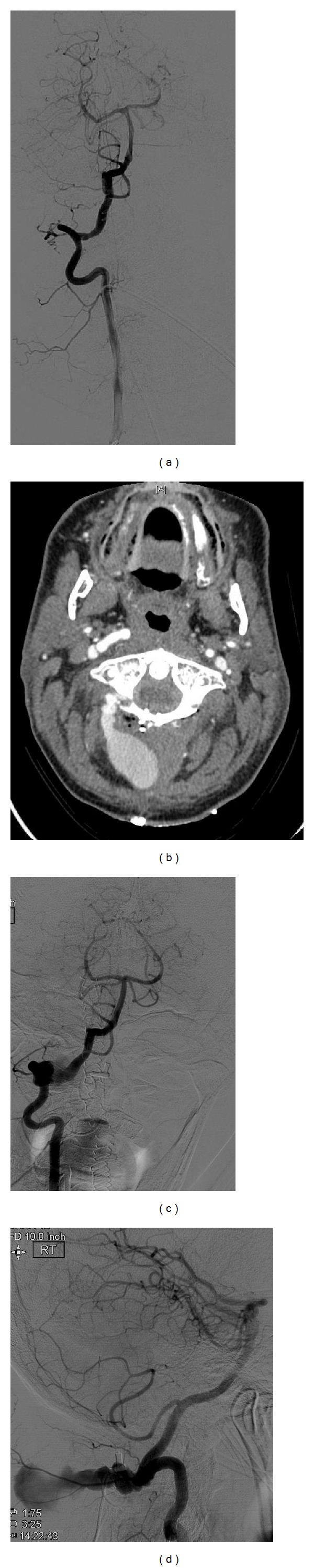
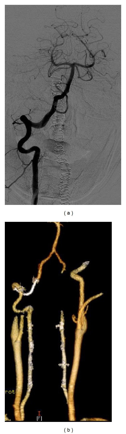
Traumatic pseudoaneurysms are uncommon and one of the most difficult lesions to treat. Traditional treatment methods have focused on parent vessel sacrifice with or without revascularization. We report the case of a patient who underwent successful treatment of an iatrogenic extracranial vertebral artery pseudoaneurysm using the Pipeline Embolization Device. A 47-year-old man sustained an inadvertent injury to the left vertebral artery during C1-C2 fixation. Subsequent imaging revealed an iatrogenic vertebral artery pseudoaneurysm. Immediate angiogram was normal. A repeat angiogram done after 3 days of the surgery revealed a vertebral artery pseudoaneurysm. He underwent aneurysm exclusion and vascular reconstruction using the Pipeline Embolization Device. Although flow-diverting stents are currently not being used for treating traumatic pseudoaneurysms, their use may be considered in such cases if active bleeding has ceased. In our case, the patient did well and the aneurysm was excluded from circulation while reconstructing the vessel wall.
Figures
References
-
- Neo M, Fujibayashi S, Miyata M, Takemoto M, Nakamura T. Vertebral artery injury during cervical spine surgery: a survey of more than 5600 operations. Spine. 2008;33(7):779–785. - PubMed
-
- Yeom JS, Buchowski JM, Park KW, Chang BS, Lee CK, Riew KD. Undetected vertebral artery groove and foramen violations during C1 lateral mass and C2 pedicle screw placement. Spine. 2008;33(25):E942–E949. - PubMed
-
- Larson PS, Reisner A, Morassutti DJ, Abdulhadi B, Harpring JE. Traumatic intracranial aneurysms. Neurosurgical Focus. 2000;8(1, article e4) - PubMed
-
- Holmes B, Harbaugh RE. Traumatic intracranial aneurysms: a contemporary review. The Journal of Trauma. 1993;35(6):855–860. - PubMed
LinkOut - more resources
Full Text Sources
Other Literature Sources
Miscellaneous
