

Effect of nicotine on the structure of cochlea of guinea pigs
- PMID: 25276475
- PMCID: PMC4178191
- DOI: 10.5115/acb.2014.47.3.162
Effect of nicotine on the structure of cochlea of guinea pigs
Abstract
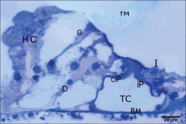
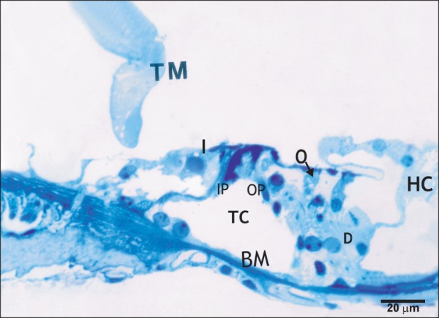
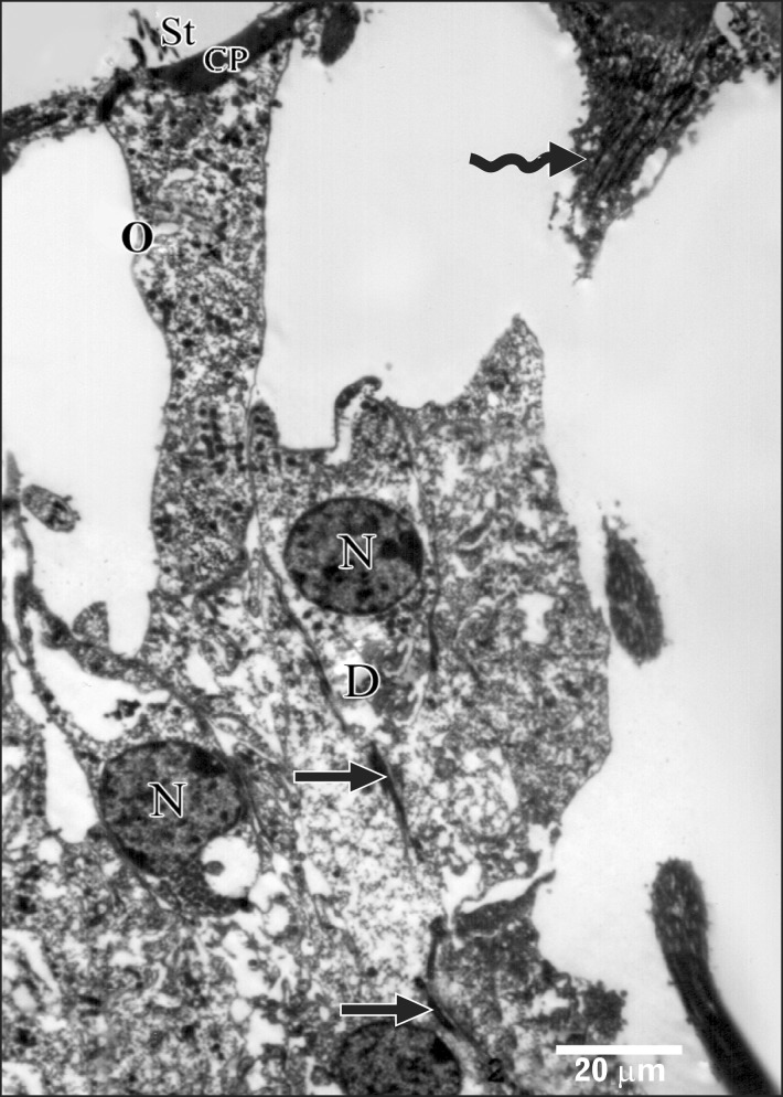
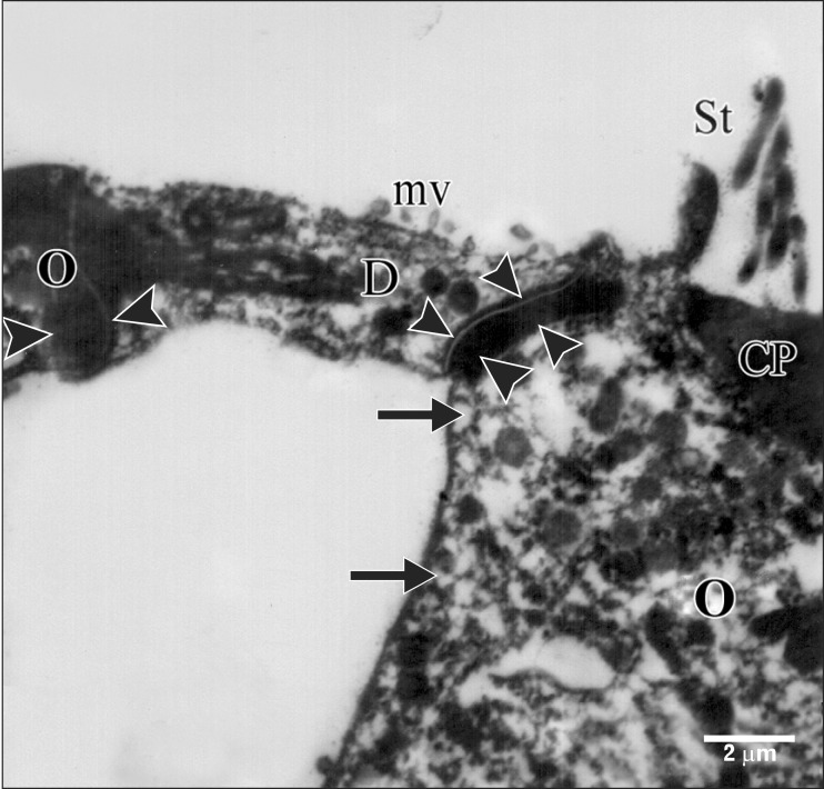
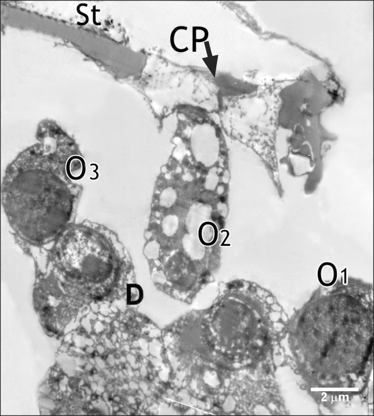
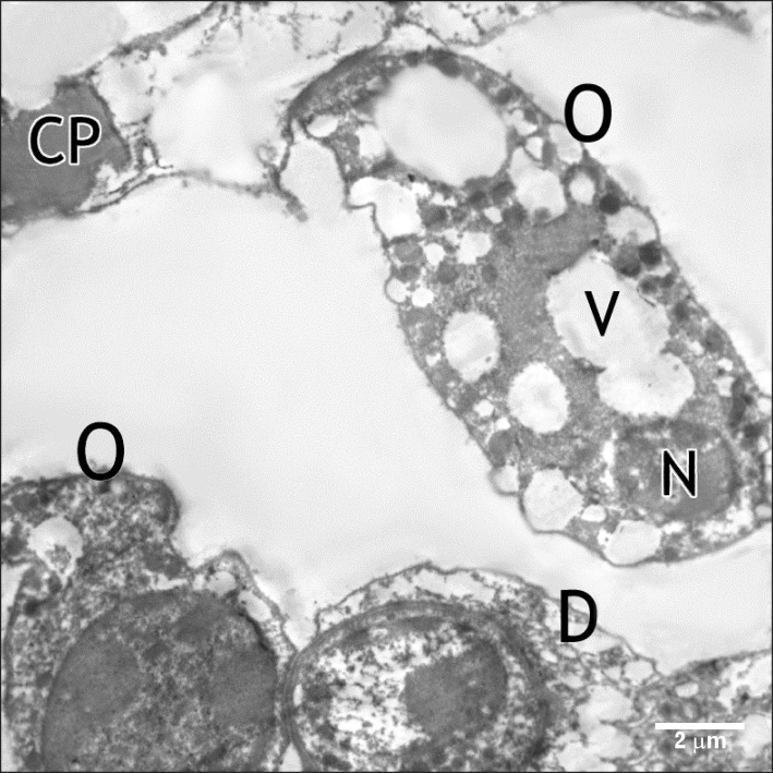
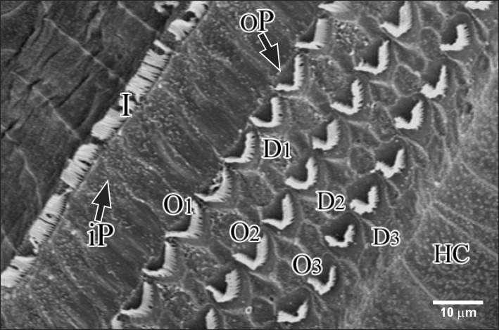
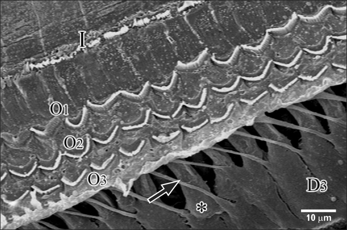
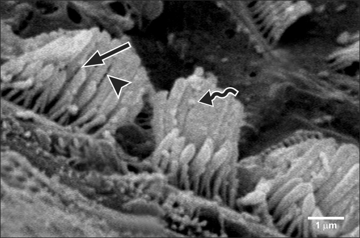
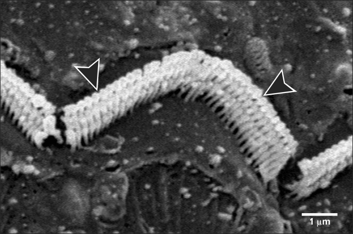
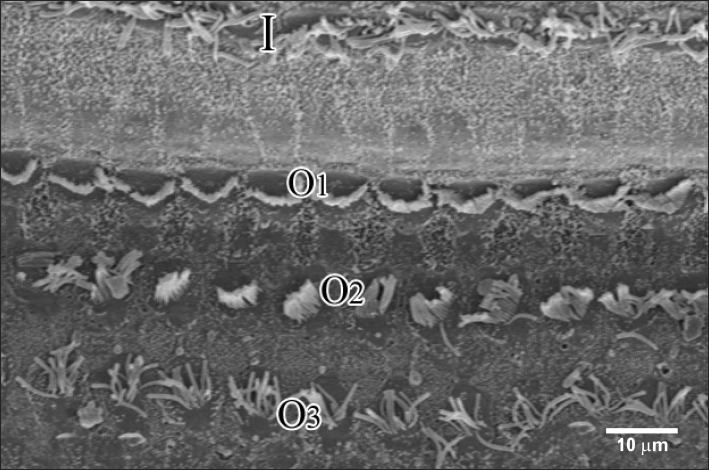
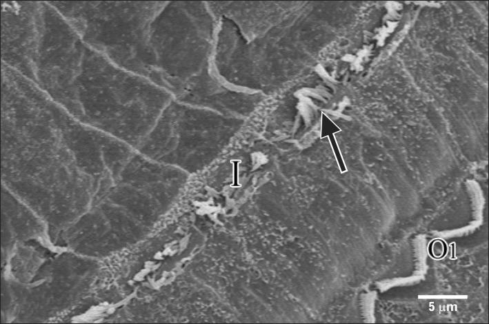
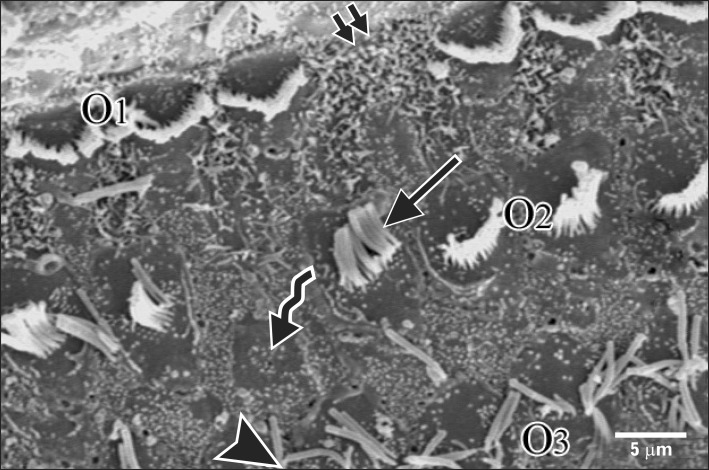
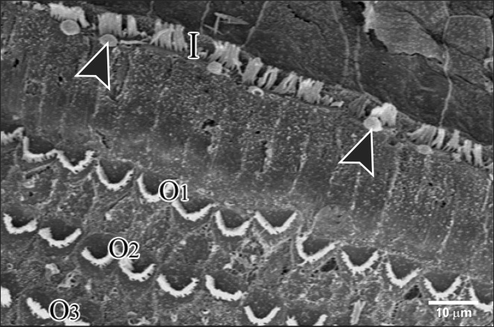
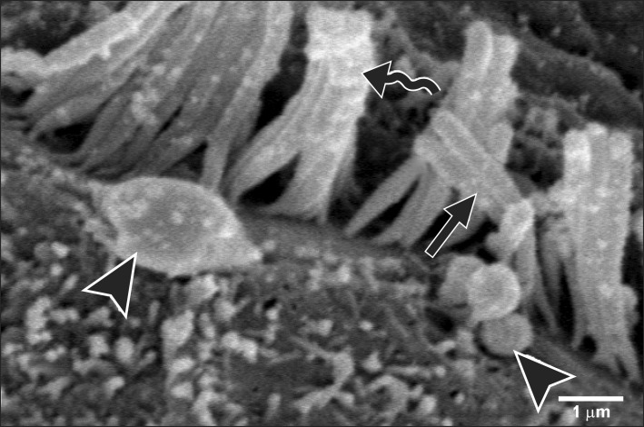
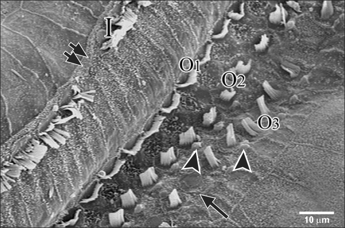
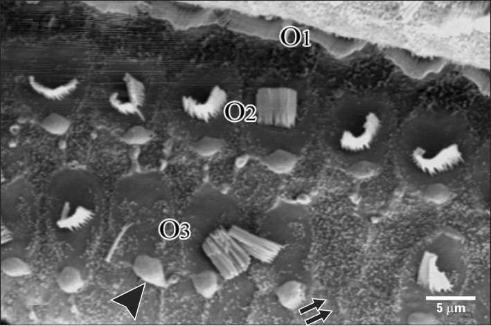
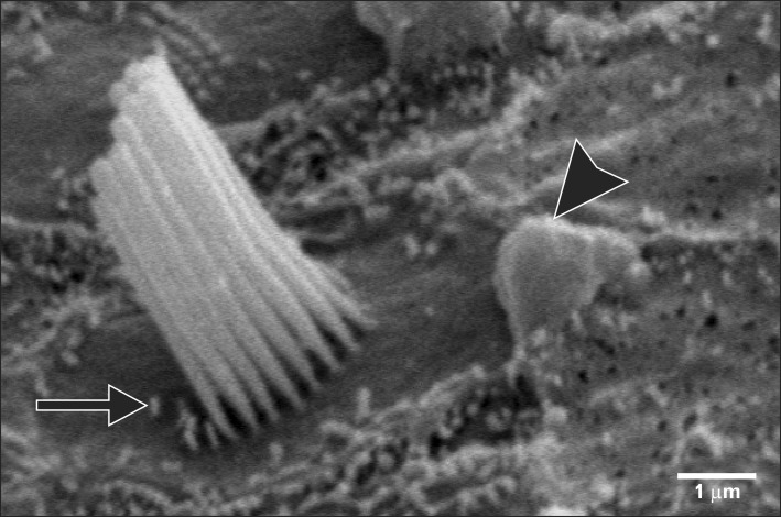
Smoking has been positively associated with hearing loss in human. However, its effect on the cochlea has not been previously evaluated. Aim of work is to investigate the effect of nicotine, which is the primary pharmacological component of tobacco, on the structure of the cochlea of adult male guinea pigs. Fifteen male guinea pigs were classified into two groups: group I (control) and group II (nicotine treated group). Group II was further subdivided into two subgroups; IIA and IIB according to the dose of nicotine (3 mg/kg and 6 mg/kg, respectively). The cochlea was harvested and processed for light microscopy, transmission electron microscopy and scanning electron microscopy. Nicotine administration induced damage of outer hair cells which were distorted in shape with vacuolated cytoplasm and heterochromatic nuclei. Topography revealed damage of the stereocilia which included disorganization, bent and limp or complete loss and expansion of the surrounding supporting cells. These changes were more pronounced in the basal turn of the cochlea and mainly involved the outer hair cells. High dose induced more damage and resulted in protrusion of the apical poles of hair cells (blebing), particularly the outer two rows. Nicotine is proved to be harmful to the cells of the cochlea, particularly the outer hair cells of the basal turn. High doses induce blebing of hair cells.
Keywords: Cochlea; Guinea pigs; Nicotine; Scanning electron microscopy.
Figures
Similar articles
-
Ultrastructural changes in the cochlea of the guinea pig after fast neutron irradiation.Otolaryngol Head Neck Surg. 1994 Apr;110(4):419-27. doi: 10.1177/019459989411000412. Otolaryngol Head Neck Surg. 1994. PMID: 8170687
-
Hair cell loss in the aged guinea pig cochlea.Acta Otolaryngol. 1999 Jan;119(1):42-7. doi: 10.1080/00016489950181918. Acta Otolaryngol. 1999. PMID: 10219383
-
Ototoxicity of tobramycin in young adult and old rats.Toxicol Appl Pharmacol. 1996 Jan;136(1):179-85. doi: 10.1006/taap.1996.0022. Toxicol Appl Pharmacol. 1996. PMID: 8560472
-
[The experimental study of the anti-damage effect of iminoethyl-lysine on noise-induced cochlea damage in guinea pig].Zhonghua Lao Dong Wei Sheng Zhi Ye Bing Za Zhi. 2002 Oct;20(5):356-8. Zhonghua Lao Dong Wei Sheng Zhi Ye Bing Za Zhi. 2002. PMID: 14694725 Chinese.
-
Early hair-cell degeneration in the extreme apex of the guinea pig cochlea.Hear Res. 1994 Sep;79(1-2):147-60. doi: 10.1016/0378-5955(94)90136-8. Hear Res. 1994. PMID: 7806477
Cited by
-
Histological study on the effect of nicotine on adult male guinea pig thin skin.Anat Cell Biol. 2017 Sep;50(3):187-199. doi: 10.5115/acb.2017.50.3.187. Epub 2017 Sep 20. Anat Cell Biol. 2017. PMID: 29043097 Free PMC article.
-
Impact of Nicotine Exposure on Hair Cell Toxicity and Embryotoxicity During Zebrafish Development.Clin Exp Otorhinolaryngol. 2018 Jun;11(2):109-117. doi: 10.21053/ceo.2017.00857. Epub 2018 Jan 6. Clin Exp Otorhinolaryngol. 2018. PMID: 29307133 Free PMC article.
-
Nicotine induced ototoxicity in rat cochlear organotypic cultures.Transl Neurosci. 2021 Oct 26;12(1):407-414. doi: 10.1515/tnsci-2020-0191. eCollection 2021 Jan 1. Transl Neurosci. 2021. PMID: 34745657 Free PMC article.
-
Maternal factors associated with smoking during gestation and consequences in newborns: Results of an 18-year study.J Clin Transl Res. 2022 Jan 3;8(1):6-19. eCollection 2022 Feb 25. J Clin Transl Res. 2022. PMID: 35097236 Free PMC article.
-
Cigarette- and snus-modified association between unprotected exposure to noise from hunting rifle caliber weapons and high frequency hearing loss. A cross-sectional study among swedish hunters.Noise Health. 2016 Nov-Dec;18(85):382-390. doi: 10.4103/1463-1741.195796. Noise Health. 2016. PMID: 27991471 Free PMC article.
References
-
- Hiremagalur B, Sabban EL. Nicotine elicits changes in expression of adrenal catecholamine biosynthetic enzymes, neuropeptide Y and immediate early genes by injection but not continuous administration. Brain Res Mol Brain Res. 1995;32:109–115. - PubMed
-
- Ingram JR, Routledge P, Rhodes J, Marshall RW, Buss DC, Evans BK, Feyerabend C, Thomas GA. Nicotine enemas for treatment of ulcerative colitis: a study of the pharmacokinetics and adverse events associated with three doses of nicotine. Aliment Pharmacol Ther. 2004;20:859–865. - PubMed
-
- McClernon FJ, Hiott FB, Westman EC, Rose JE, Levin ED. Transdermal nicotine attenuates depression symptoms in nonsmokers: a double-blind, placebo-controlled trial. Psychopharmacology (Berl) 2006;189:125–133. - PubMed
LinkOut - more resources
Full Text Sources
Other Literature Sources