

Intracranial injectable tumor model: technical advancements
- PMID: 25276597
- PMCID: PMC4176537
- DOI: 10.1055/s-0034-1368148
Intracranial injectable tumor model: technical advancements
Abstract
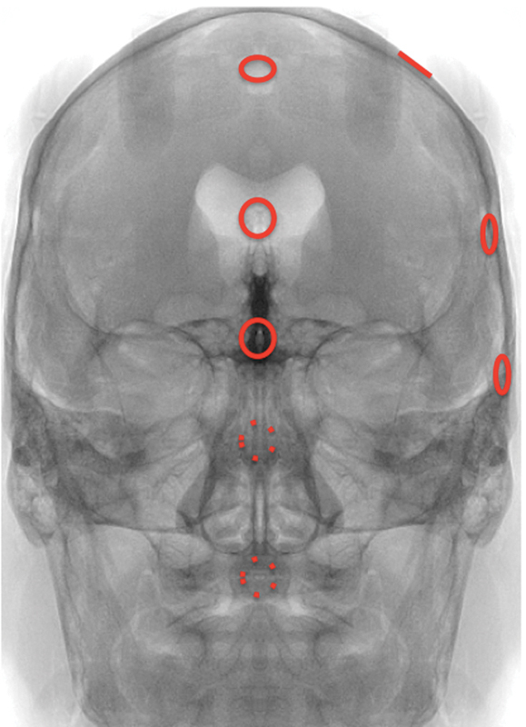
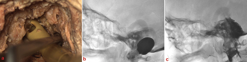
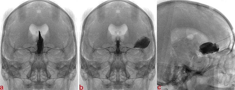
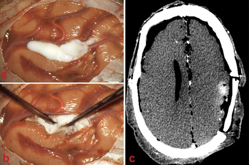
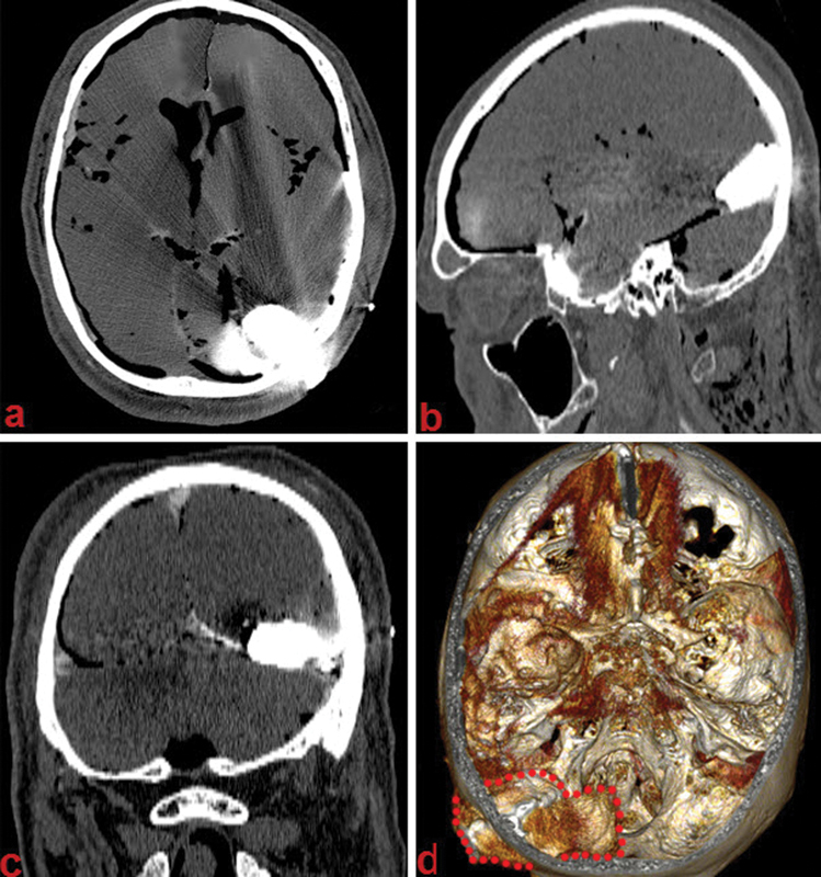
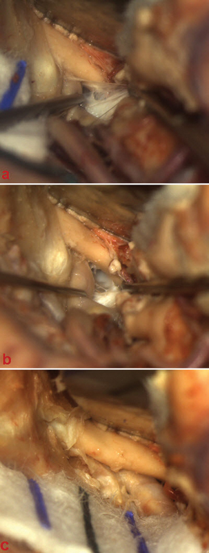
Background and Objectives Few simulation models are available that provide neurosurgical trainees with the challenge of distorted skull base anatomy despite increasing importance in the acquisition of safe microsurgical and endoscopic techniques. We have previously reported a unique training model for skull base neurosurgery where a polymer is injected into a cadaveric head where it solidifies to mimic a skull base tumor for resection. This model, however, required injection of the polymer under direct surgical vision via a complicated alternative approach to that being studied, prohibiting its uptake in many neurosurgical laboratories. Conclusion We report our updated skull base tumor model that is contrast-enhanced and may be easily and reliably injected under fluoroscopic guidance. We have identified a map of burr holes and injection corridors available to place tumor at various intracranial sites. Additionally, the updated tumor model allows for the creation of mass effect, and we detail the variation of polymer preparation to mimic different tumor properties. These advancements will increase the practicality of the tumor model and ideally influence neurosurgical standards of training.
Keywords: microsurgery; neurosurgical training; skull base; tumor model.
Figures
References
-
- Gragnaniello C, Nader R, van Doormaal T. et al. Skull base tumor model. J Neurosurg. 2010;113(5):1106–1111. - PubMed
-
- Sanan A Abdel Aziz K M Janjua R M van Loveren H R Keller J T Colored silicone injection for use in neurosurgical dissections: anatomic technical note Neurosurgery 19994551267–1271., discussion 1271–1274 - PubMed
-
- Zhao J C Chen C Rosenblatt S S et al. Imaging the cerebrovascular tree in the cadaveric head for planning surgical strategy Neurosurgery 20025151222–1227.; discussion 1227–1228 - PubMed
-
- Cunha e Sá M. Additional competence training charter in neurosurgical oncology. Acta Neurochir (Wien) 2010;152(6):1095–1098. - PubMed
-
- Abe M Tabuchi K Goto M Uchino A Model-based surgical planning and simulation of cranial base surgery Neurol Med Chir (Tokyo) 19983811746–750.; discussion 750–751 - PubMed
LinkOut - more resources
Full Text Sources
Other Literature Sources
