

Electrophysiological correlates of the BOLD signal for EEG-informed fMRI
- PMID: 25277370
- PMCID: PMC4280889
- DOI: 10.1002/hbm.22623
Electrophysiological correlates of the BOLD signal for EEG-informed fMRI
Abstract
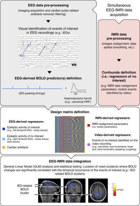
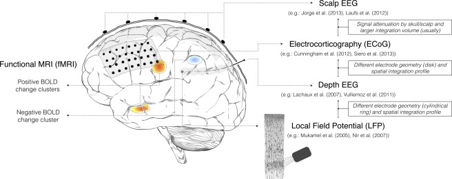
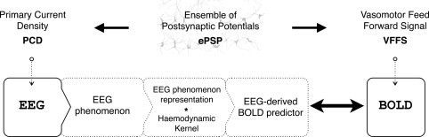
Electroencephalography (EEG) and functional magnetic resonance imaging (fMRI) are important tools in cognitive and clinical neuroscience. Combined EEG-fMRI has been shown to help to characterise brain networks involved in epileptic activity, as well as in different sensory, motor and cognitive functions. A good understanding of the electrophysiological correlates of the blood oxygen level-dependent (BOLD) signal is necessary to interpret fMRI maps, particularly when obtained in combination with EEG. We review the current understanding of electrophysiological-haemodynamic correlates, during different types of brain activity. We start by describing the basic mechanisms underlying EEG and BOLD signals and proceed by reviewing EEG-informed fMRI studies using fMRI to map specific EEG phenomena over the entire brain (EEG-fMRI mapping), or exploring a range of EEG-derived quantities to determine which best explain colocalised BOLD fluctuations (local EEG-fMRI coupling). While reviewing studies of different forms of brain activity (epileptic and nonepileptic spontaneous activity; cognitive, sensory and motor functions), a significant attention is given to epilepsy because the investigation of its haemodynamic correlates is the most common application of EEG-informed fMRI. Our review is focused on EEG-informed fMRI, an asymmetric approach of data integration. We give special attention to the invasiveness of electrophysiological measurements and the simultaneity of multimodal acquisitions because these methodological aspects determine the nature of the conclusions that can be drawn from EEG-informed fMRI studies. We emphasise the advantages of, and need for, simultaneous intracranial EEG-fMRI studies in humans, which recently became available and hold great potential to improve our understanding of the electrophysiological correlates of BOLD fluctuations.
Keywords: correlation; coupling; electrophysiology; functional magnetic resonance imaging; human brain.
© 2014 The Authors Human Brain Mapping Published by Wiley Periodicals, Inc.
Figures
References
-
- Aguirre GK, Zarahn E, D'Esposito M (1998): The variability of human, BOLD hemodynamic responses. Neuroimage 8:360–369. - PubMed
-
- Al‐Asmi A, Benar CG, Gross DW, Khani YA, Andermann F, Pike B, Dubeau F, Gotman J (2003): fMRI activation in continuous and spike‐triggered EEG‐fMRI studies of epileptic spikes. Epilepsia 44:1328–1339. - PubMed
-
- Allen PJ, Josephs O, Turner R (2000): A method for removing imaging artifact from continuous EEG recorded during functional MRI. Neuroimage 12:230–239. - PubMed
-
- Attwell D, Iadecola C (2002): The neural basis of functional brain imaging signals. Trends Neurosci 25:621–625. - PubMed
Publication types
MeSH terms
Substances
Grants and funding
LinkOut - more resources
Full Text Sources
Other Literature Sources
