Review
. 2014 Nov:468-470:565-580.
doi: 10.1016/j.virol.2014.09.009. Epub 2014 Sep 29.

Phosphatidylserine receptors: enhancers of enveloped virus entry and infection

Affiliations
Review

Phosphatidylserine receptors: enhancers of enveloped virus entry and infection

Sven Moller-Tank et al. Virology. 2014 Nov.

Abstract

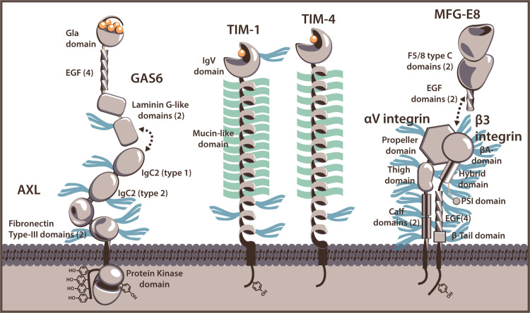
A variety of both RNA and DNA viruses envelop their capsids in a lipid bilayer. One of the more recently appreciated benefits this envelope is incorporation of phosphatidylserine (PtdSer). Surface exposure of PtdSer disguises viruses as apoptotic bodies; tricking cells into engulfing virions. This mechanism is termed apoptotic mimicry. Several PtdSer receptors have been identified to enhance virus entry and we have termed this group of proteins PtdSer-mediated virus entry enhancing receptors or PVEERs. These receptors enhance entry of a range of enveloped viruses. Internalization of virions by PVEERs provides a broad mechanism of entry with little investment by the virus itself. PVEERs may allow some viruses to attach to cells, thereby making viral glycoprotein/cellular receptor interactions more probable. Alternatively, other viruses may rely entirely on PVEERs for internalization into endosomes. This review provides an overview of PtdSer receptors that serve as PVEERs and the biology behind virion/PVEER interaction.

Keywords: Alphavirus; Axl; Baculovirus; Enveloped virus; Filovirus; Flavivirus; Integrin αvβ3; Integrin αvβ5; MFG-E8; Mer; PVEER; Phosphatidylserine; Phosphatidylserine receptor; Receptors; TAM; TIM-1; TIM-4; Tyro3; Virus entry.


Figures

Fig. 1
Fig. 1
PtdSer-binding receptors that function as PVEERs. Cartoon representations of Gas 6/Axl (representatives of TAM ligand and receptor kinases), TIM-1, TIM-4, and MFG-E8/ αvβ3 integrin are displayed. Estimations of N-linked glycan sites are represented as blue tridents and O-link glycoslyations (TIM-1 and TIM-4 only) are shown as green lines. Domains for which calcium-binding is necessary for interaction with PtdSer are indicated with calcium ions (orange spheres). Gas6 and Axl interact through their respective laminin G-like and IgC2 domains. MFG-E8 contains an Arg-Gly-Asp (RGD) motif in the second EGF domain that likely binds at the interface of the αvβ3/5 integrin complex (Xiong et al., 2002).
Fig. 2
Fig. 2
Structures of PtdSer-binding domains. Representative renders of the PtdSer-binding domains of TIM-1/-3/-4, CD300a, RAGE, BAI-1, Gas6/Protein S, Annexin V, MFG-E8, and Stabilin-1/2 are shown. PtdSer-like ligands (red) and calcium ions (orange) are also displayed for structures with which they were solved. For domains that do not have solved structures, equivalent domains from other proteins are shown. The IgV domain of TIM-1/-3/-4 is represented by the mTIM-4 IgV domain (2OR8) (Santiago et al., 2007). The human CD300a IgV domain crystal structure (Dimasi et al., 2007) is shown with residues hypothesized for interaction (Simhadri et al., 2012). The region of the RAGE IgV domain (3O3U) that corresponds to the binding pockets of CD300a and TIM-1 IgV domains is shown for comparison (Park et al., 2010); however, this domain is only hypothesized to interact with PtdSer and no residues have been implicated (Friggeri et al., 2011). The thrombospondin type-1 repeats (TSRs) of BAI1 are represented by TSR domain 3 of human thrombospondin-1 (3R6B) (Klenotic et al., 2011). Gas6 binds PtdSer via a Gla domain, represented by that of bovine prothrombin (1NL2) with residues identified that interact with PtdSer (red) (Huang et al., 2003). For AnxV, the structure of one annexin repeat domain from rat Anx V is shown (1A8A) with residues identified that interact with PtdSer (red) (Swairjo et al., 1995). The C2 domain of bovine MFG-E8 binds to PtdSer (3BN6) (Shao et al., 2008) and while the structure was not crystallized with PtdSer, key residues involved in interaction have been determined experimentally (Ye et al., 2013). An EGF-like domain of stabilin-1 or -2 is represented by that from human heregulin alpha (1HRE) (Nagata et al., 1994).
Fig. 3
Fig. 3
PtdSer-binding receptors that do not function as PVEERs. Cartoon representations of Stabilin-1, BAI1, CD300a, RAGE, and TIM-3 are shown. Estimations of N-linked glycan sites are indicated with blue tridents and O-link glycosylation sites (TIM-3 only) are indicated with a green line. The binding pocket of the RAGE IgV domain is only hypothesized, as indicated by the presence of a dotted line. Domains for which calcium-binding is necessary for interaction with PtdSer are shown with calcium ions (orange spheres).

References

    1. Abraham J, Kwong JA, Albarino CG, Lu JG, Radoshitzky SR, Salazar-Bravo J, Farzan M, Spiropoulou CF, Choe H. Host-species transferrin receptor 1 orthologs are cellular receptors for nonpathogenic new world clade B arenaviruses. PLoS Pathog. 2009;5:e1000358. - PMC - PubMed
    1. Acosta EG, Castilla V, Damonte EB. Alternative infectious entry pathways for dengue virus serotypes into mammalian cells. Cell. Microbiol. 2009;11:1533–1549. - PMC - PubMed
    1. Adu-Gyamfi E, Soni SP, Xue Y, Digman MA, Gratton E, Stahelin RV. The Ebola virus matrix protein penetrates into the plasma membrane: a key step in viral protein 40 (VP40) oligomerization and viral egress. J. Biol. Chem. 2013;288:5779–5789. - PMC - PubMed
    1. Ajay AK, Kim T-M, Ramirez-Gonzalez V, Park PJ, Frank DA, Vaidya VS. A bioinformatics approach identifies signal transducer and activator of ranscription-3 and checkpoint kinase 1 as upstream regulators of kidney injury molecule-1 after kidney injury. J. Am. Soc.Nephrol. 2014;25:105–118. - PMC - PubMed
    1. Akhtar J, Shukla D. Viral entry mechanisms: cellular and viral mediators of herpes simplex virus entry. FEBS J. 2009;276:7228–7236. - PMC - PubMed

MeSH terms