Review
. 2015 Feb:146:104-19.
doi: 10.1016/j.pharmthera.2014.09.009. Epub 2014 Sep 30.

Rabbit models for the study of human atherosclerosis: from pathophysiological mechanisms to translational medicine

Affiliations
Review

Rabbit models for the study of human atherosclerosis: from pathophysiological mechanisms to translational medicine

Jianglin Fan et al. Pharmacol Ther. 2015 Feb.

Abstract

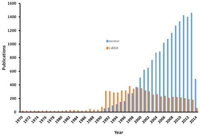
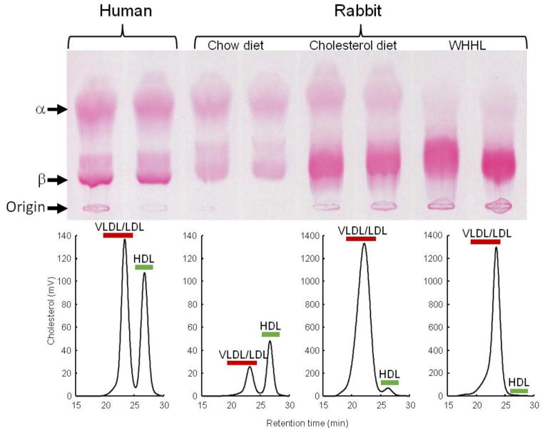
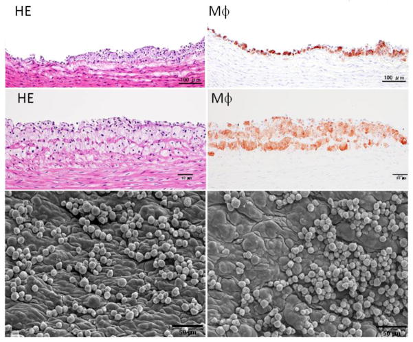
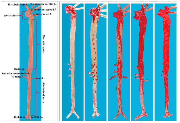
Laboratory animal models play an important role in the study of human diseases. Using appropriate animals is critical not only for basic research but also for the development of therapeutics and diagnostic tools. Rabbits are widely used for the study of human atherosclerosis. Because rabbits have a unique feature of lipoprotein metabolism (like humans but unlike rodents) and are sensitive to a cholesterol diet, rabbit models have not only provided many insights into the pathogenesis and development of human atherosclerosis but also made a great contribution to translational research. In fact, rabbit was the first animal model used for studying human atherosclerosis, more than a century ago. Currently, three types of rabbit model are commonly used for the study of human atherosclerosis and lipid metabolism: (1) cholesterol-fed rabbits, (2) Watanabe heritable hyperlipidemic rabbits, analogous to human familial hypercholesterolemia due to genetic deficiency of LDL receptors, and (3) genetically modified (transgenic and knock-out) rabbits. Despite their importance, compared with the mouse, the most widely used laboratory animal model nowadays, the use of rabbit models is still limited. In this review, we focus on the features of rabbit lipoprotein metabolism and pathology of atherosclerotic lesions that make it the optimal model for human atherosclerotic disease, especially for the translational medicine. For the sake of clarity, the review is not an attempt to be completely inclusive, but instead attempts to summarize substantial information concisely and provide a guideline for experiments using rabbits.

Keywords: Atherosclerosis; Experimental animal models; Hypercholesterolemia; Transgenic rabbits; Translational medicine.


Figures

Fig. 1
Fig. 1
Total number of research articles reporting the use of rabbits and mice in research on atherosclerosis from 1970 to 2014 (Source: Web of Science).
Fig. 2
Fig. 2
Lipoprotein profiles of wild-type rabbits on a normal chow diet and cholesterol-fed, as well as WHHL rabbits, compared with human. Agarose gel electrophoresis of plasma lipoproteins (upper panel) and FPLC analysis of lipoproteins (lower panel).
Fig. 3
Fig. 3
Early pathological changes of aortic surface of cholesterol-fed rabbits. Two representative lesions are shown (the top and middle panels) and were subjected to either hematoxylin and eosin (HE)staining (left) or immunohistochemical staining with RAM11 antibody against rabbit macrophage (Mϕ) (right). Aortic lesions are also visible under a scanning electron microscope (lower panels). Many monocytes either singly or in clusters adhere to the endothelial cells of the aorta.
Fig. 4
Fig. 4
Gross lesions of aortic atherosclerosis in cholesterol-fed rabbits. The normal aorta is cut open and shown as a reference (left) to illustrate rabbit aortic tree anatomy. Five aortas stained by Sudan IV show different degrees of aortic lesions (red areas stained with Sudan IV).
Fig. 5
Fig. 5
Representative fatty streaks of aortic atherosclerosis in cholesterol-fed rabbits. The lesions are composed of accumulated macrophage-derived foam cells in the center and smooth muscle cells on the top. Arrowhead indicates the necrotic core. The specimen is stained with HE and elastic van Gieson (EVG) or immunohistochemically stained with RAM11 antibody against rabbit macrophage (Mϕ) and HHF35 antibody for smooth muscle cells (SMC).
Fig. 6
Fig. 6
Advanced lesions of aortic atherosclerosis in cholesterol-fed rabbits. The lesions contain a small number of macrophages and calcium deposition can be seen, as indicated by arrowheads.
Fig. 7
Fig. 7
“Fibrotic lesions” of aortic atherosclerosis in cholesterol-fed rabbits. This lesion is less frequent and characterized by fibrosis and smooth muscle cell accumulation with fewer macrophages.
Fig. 8
Fig. 8
A typical foam cell-rich lesion of aortic atherosclerosis in cholesterol-fed rabbits with extremely high hypercholesterolemia. The lesions are almost completely composed of foam cells, which can be stained with RAM11 antibody.
Fig. 9
Fig. 9
Typical coronary atherosclerosis in cholesterol-fed rabbits. The lesions are mainly composed of smooth muscle cells and occupy about 50% of the lumen.
Fig. 10
Fig. 10. Different features of aortic atherosclerotic lesions in WHHL rabbits
A. The foam cell-rich lesion is composed of macrophages and covered by a few smooth muscle cells on the top. B. A fibrous plaque contains a lipid core (arrowheads) in the center. C. Another fibrous plaque contains a large necrotic core (arrowheads) in the center, which is covered by a thin fibrous cap. D. Advanced lesions show marked calcification defined by dashed lines.
Fig. 11
Fig. 11. Typical coronary atherosclerosis in WHHL rabbits
A. A typical fatty streak with many macrophages leads to stenosis. B. An advanced lesion with calcification (arrowheads) shows remarkable stenosis.
Fig. 11
Fig. 11. Typical coronary atherosclerosis in WHHL rabbits
A. A typical fatty streak with many macrophages leads to stenosis. B. An advanced lesion with calcification (arrowheads) shows remarkable stenosis.
Fig. 12
Fig. 12
Schematic illustration of zinc finger, TALE and RNA-guided nucleases for genome editing.

References

    1. Aikawa M, Voglic SJ, Sugiyama S, Rabkin E, Taubman MB, Fallon JT, Libby P. Dietary lipid lowering reduces tissue factor expression in rabbit atheroma. Circulation. 1999;100:1215–1222. - PubMed
    1. Badimon JJ, Badimon L, Fuster V. Regression of atherosclerotic lesions by high density lipoprotein plasma fraction in the cholesterol-fed rabbit. J Clin Invest. 1990;85:1234–1241. - PMC - PubMed
    1. Badimon JJ, Badimon L, Galvez A, Dische R, Fuster V. High density lipoprotein plasma fractions inhibit aortic fatty streaks in cholesterol-fed rabbits. Lab Invest. 1989;60:455–461. - PubMed
    1. Besterman EM. Experimental coronary atherosclerosis in rabbits. Atherosclerosis. 1970;12:75–83. - PubMed
    1. Bocan TM, Mueller SB, Mazur MJ, Uhlendorf PD, Brown EQ, Kieft KA. The relationship between the degree of dietary-induced hypercholesterolemia in the rabbit and atherosclerotic lesion formation. Atherosclerosis. 1993;102:9–22. - PubMed

Publication types