. 2014 Nov 14;289(46):31805-31817.
doi: 10.1074/jbc.M113.541219. Epub 2014 Oct 2.

The E3 ubiquitin ligase MID1 catalyzes ubiquitination and cleavage of Fu

Affiliations

The E3 ubiquitin ligase MID1 catalyzes ubiquitination and cleavage of Fu

Susann Schweiger et al. J Biol Chem. .

Abstract

SHH (Sonic Hedgehog)-GLI signaling plays an important role during embryogenesis and in tumorigenesis. The survival and growth of several types of cancer depend on autonomously activated SHH-GLI signaling. A protein complex containing the ubiquitin ligase MID1 and protein phosphatase 2A regulates the nuclear localization and transcriptional activity of GLI3, a transcriptional effector molecule of SHH, in cancer cell lines with autonomously activated SHH signaling. However, the exact molecular mechanisms that mediate the interaction between MID1 and GLI3 remained unknown. Here, we show that MID1 catalyzes the ubiquitination and proteasomal cleavage of the GLI3 regulator Fu. Our data suggest that Fu ubiquitination and cleavage is one of the key elements connecting the MID1-PP2A protein complex with GLI3 activity control.

Keywords: Cancer Biology; Fu; GLI3; MID1; Proteasomal Cleavage; Proteasome; Protein Phosphatase 2 (PP2A); Signal transduction; Ubiquitination.


Figures

FIGURE 1.
FIGURE 1.
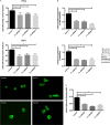
Subcellular localization and transcriptional activity of GLI3 after Fu knockdown. A, subcellular distribution of GFP-GLI3 in HeLa (top panel) or U373MG (middle panel) cells after co-transfection with either nonsilencing control siRNAs or three different Fu-specific siRNAs. GFP signal distribution in individual cells occurred in three patterns: exclusively nuclear fluorescence, even staining throughout cytosol and nucleus, or predominantly cytosolic fluorescence. Transfected cells were randomly chosen, counted, and classified in one of the three groups. The data shown represent percentages of cells showing strictly nuclear GFP-GLI3 localization (means ± S.D. scored per group from three independent experiments of 100 cells each). *, p < 0.05. Bottom panel, representative pictures of cells showing GFPGLI3 localization after transfection with nonsilencing control siRNAs or three different Fu-specific siRNAs. B, relative Cyclin D1 (upper panel) or Fu (lower panel) mRNA amounts in HeLa cells transfected with either nonsilencing control siRNAs or three different Fu-specific siRNAs as measured by real time PCR (lower panel, left) and Western blot (lower panel, right) using Fu- or tubulin-specific antibodies. Left, columns represent mean values of four samples measured in parallel ± S.D. GAPDH was used for normalization. *, p < 0.05. C, GLI3 reporter assay. Firefly luciferase under the control of eight GLI-binding sites was co-transfected with three different Fu-specific siRNAs or control-siRNAs. As an internal transfection control, Renilla luciferase was included and used for normalization. The columns show relative firefly luciferase signals from four samples ± S.D. *, p < 0.05.
FIGURE 2.
FIGURE 2.
Interaction of Fu with the ubiquitin ligase MID1. A, co-immunoprecipitation of FLAG-MID1 and Fu-V5. Immunoprecipitations from HeLa cell lysates overexpressing FLAG-MID1 and Fu-V5 were performed using anti-V5 antibodies, anti-FLAG-M2-agarose, or nonspecific IgGs as negative control. Immunoprecipitates were analyzed by Western blot. FLAG-MID1 (lower panel) and Fu-V5 (upper panel) were detected using the respective antibodies. The full-length FLAG-MID1 and Fu-V5 bands, as well as shorter Fu-V5 fragments and a high molecular weight Fu-V5 smear are depicted. B, in vitro ubiquitination of HA-Fu. HA-Fu was incubated with an E1 enzyme, with a panel of 11 different E2 enzymes (UbcH1, UbcH2, UbcH3, UbcH5a, UbcH5b, UbcH5c, UbcH6, UbcH7, UbcH8, UbcH10, and Ubc13), and with (right panels) or without (left panels) the E3 enzyme MID1 and analyzed on a Western blot. The binding of biotinylated ubiquitin to HA-Fu was visualized using HRP-streptavidin (upper panels). To show equal loading of HA-Fu to each reaction, the same membranes were incubated with anti-HA antibodies (lower panels). The full-length HA-Fu bands, as well as shorter HA-Fu fragments (asterisk), are depicted.
FIGURE 3.
FIGURE 3.
A, immunoprecipitation of HA-Fu conjugated to FLAG ubiquitin. HA-Fu was co-expressed with FLAG ubiquitin with or without treatment with the proteasome inhibitor MG-132 and purified by immunoprecipitation using HA antibodies. The incorporation of ubiquitin was then detected on Western blots using FLAG antibodies (upper panel). To show the loading of HA-Fu to each lane, the same membranes were incubated with anti-Fu antibodies (lower panel). B, in vitro ubiquitination of HA-Fu. HA-Fu was incubated with an E1 enzyme, with the E2 enzyme UbcH5b, and with (+MID1) or without (−MID1) the E3 enzyme MID1 and analyzed on a Western blot. In this reaction, either WT ubiquitin or ubiquitin proteins in which all lysines have been mutated to arginine (KO), in which all lysines but Lys48 have been mutated to arginine (Lys48), or in which all lysines but Lys63 have been mutated to arginine (Lys63) were applied. The binding of biotinylated ubiquitin to HA-Fu was visualized using HRP-streptavidin (upper panel). To show equal loading of HA-Fu to each reaction, the same membranes were incubated with anti-HA antibodies (lower panel). C, in vitro ubiquitination of HA-Fu. HA-Fu was incubated either without or with a mixture of an E1 enzyme, the E2 enzyme UbcH5b, the E3 enzyme MID1, and methylated ubiquitin and analyzed on a Western blot. The binding of methylated ubiquitin to HA-Fu induced a shift in the molecular weight, which was detected with HA antibodies. D, immunoprecipitation of Fu-V5 conjugated to FLAG ubiquitin. Full-length Fu-V5 or a deletion construct lacking amino acids 8–100 (Δ8–100) was co-expressed with FLAG ubiquitin. Ubiquitinated proteins were purified by immunoprecipitation using anti-FLAG beads. Ubiquitinated Fu was then detected on Western blots using V5 antibodies. E, in vitro ubiquitination of Fu-V5. Fu-V5 was incubated either without or with a mixture of an E1 enzyme, the E2 enzyme UbcH5b, the E3 enzyme MID1, and different ubiquitin mutants in which either all (KO) or all but one lysine have been mutated. Samples were analyzed on a Western blot on which ubiquitin (upper panel) or Fu-V5 (lower panel) was detected. F, immunoprecipitation of Fu-V5 conjugated to FLAG ubiquitin. Full-length Fu-V5 was co-expressed with either WT FLAG ubiquitin or FLAG ubiquitin in which all lysines apart from Lys6 were mutated. Ubiquitinated proteins were purified by immunoprecipitation using anti-FLAG beads. Ubiquitinated Fu was then detected on Western blots using V5 antibodies. G, in vitro ubiquitination of His-Fu with an E1 enzyme, the E2 enzyme UbcH5b, and the E3 enzyme MID1 as described for Fig. 2B. Either full-length MID1 (MID1) or a MID1 deletion construct missing the first 196 amino acids (including the RING finger domain; ΔRING) were used. To show equal loading of His-Fu to each reaction, the same membranes were incubated with anti-Fu antibodies (lower panel). H, immunoprecipitation of Fu-V5 conjugated to HA ubiquitin. Fu-V5 and HA ubiquitin were co-transfected with either nonsilencing control siRNAs or two different MID1-specific siRNAs (MID1si2 and MID1si4). Left panel, Fu-V5 was purified by immunoprecipitation with V5 antibodies (IP:V5). Pulldowns with unspecific IgGs were used as negative controls (IP:IgG). The immunoprecipitates were analyzed on Western blots detecting the Fu ubiquitination with anti-HA antibodies (upper panel). To visualize loading of Fu-V5 to each lane, the same membranes were incubated with anti-V5 antibodies (lower panel). Right panel, the efficiency of the MID1 knockdown procedure was monitored on Western blots using MID1-specific antibodies (upper panel) or GAPDH antibodies as loading control (lower panel).
FIGURE 4.
FIGURE 4.
A, identification of different Fu isoforms by mass spectrometry. Right panel, immunoprecipitated Fu-V5 was separated on an SDS gel stained by Coomassie. The bands marked by arrows were identified as Fu isoforms by mass spectrometry; the band marked by asterisk was identified as Hsp90. Left panel, an aliquot of the same immunoprecipitate was analyzed on a Western blot using anti-V5 antibodies. B, detection of different Fu isoforms. Lysates of cells expressing Fu-V5 (left panel) or cells expressing HA-Fu (right panel) were analyzed on Western blot. The different Fu protein isoforms were detected with anti-V5 (left panel) or anti-HA (right panel) antibodies, respectively. C, homodimerization of Fu. HA-Fu and V5-Fu were co-expressed in HeLa cells and immunoprecipitated with anti-V5 antibodies or nonspecific IgGs as negative control. HA-Fu (lower panel) and Fu-V5 (upper panel) were visualized on Western blots with V5 or HA antibodies, respectively. D, lysates of nontransfected HeLa cells (right lane) or HeLa cells transfected with Fu-V5 (left lane) were analyzed on Western blots using the affinity-purified Fu antibody. E, immunoprecipitation of FLAG ubiquitin and detection of ubiquitin-coupled endogenous Fu. HeLa cells were transfected with either WT FLAG ubiquitin or ubiquitin proteins in which all lysines have been mutated to arginine (KO) or in which either Lys48 or Lys63 have been mutated to arginine (K48R or K63R). All ubiquitinated proteins from the lysates of those cells were isolated by immunoprecipitation using anti-FLAG-agarose or mouse IgG-agarose as negative control. Ubiquitinated endogenous Fu was then detected on Western blots using anti-Fu antibodies. F, immunoprecipitation of FLAG ubiquitin and detection of ubiquitin-coupled endogenous Fu as in E. Prior to immunoprecipitation, cells were incubated with or without the proteasome inhibitor MG-132. G, co-expression of HA ubiquitin and FLAG-MID1 wild type or FLAG-MID1 lacking amino acids 1–196 (ΔRING). Ubiquitinated proteins were purified by HA immunoprecipitation, and the relative ubiquitination of Fu was determined by Western blot analysis using Fu antibodies. H, stabilization of endogenous Fu after MID1 knockdown. Lysates of cells transfected with nonsilencing control siRNAs (left lane) or MID1-specific siRNAs (right lane) were analyzed on Western blot. Full-length endogenous Fu was detected with anti-Fu antibodies. Tubulin was detected on the same membranes as loading control.
FIGURE 5.
FIGURE 5.
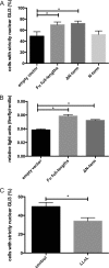
Subcellular localization and transcriptional activity of GLI3 after Fu-overexpression. A, subcellular distribution of GFP-GLI3 in HeLa cells after co-transfection with either empty vector (left columns), full-length Fu, a Fu construct lacking the N-terminal kinase domain (ΔN-term), or a construct containing the isolated Fu kinase domain (N-term, right columns). GFP-GLI3 signal distribution was quantified as described for Fig. 1A. Means ± S.D. scored per group from three independent experiments of 100 cells each are shown. *, p < 0.05. B, GLI3 reporter assay. Firefly luciferase under the control of eight GLI-binding sites was co-transfected with empty vector (left column), full-length Fu (middle column), or ΔN-term (right column). As an internal transfection control, Renilla luciferase was included and used for normalization. Columns show relative firefly luciferase signals from four samples ± S.D. *, p < 0.05. C, subcellular distribution of GFP-GLI3 in HeLa cells that were treated with or without the proteasome inhibitor N-acetyl-l-leucinyl-l-leucinal-l-norleucinal (LLnL). The data were analyzed and are presented as described for Fig. 1A. Means ± S.D. scored per group from three independent experiments of 100 cells each are shown. *, p < 0.05.
FIGURE 6.
FIGURE 6.
Hypothetical model. MID1 catalyzes the ubiquitination of Fu, thereby promoting proteasomal cleavage of Fu. The C-terminal fragment of Fu promotes nuclear localization of GLI3 and activates its target genes. Like MID1 and Fu knockdown, the inhibition of the proteasome leads to cytosolic retention of GLI3.

References

    1. Nybakken K., Perrimon N. (2002) Hedgehog signal transduction: recent findings. Curr. Opin. Genet. Dev. 12, 503–511 - PubMed
    1. Ingham P. W., McMahon A. P. (2001) Hedgehog signaling in animal development: paradigms and principles. Genes Dev. 15, 3059–3087 - PubMed
    1. Villavicencio E. H., Walterhouse D. O., Iannaccone P. M. (2000) The sonic hedgehog-patched-gli pathway in human development and disease. Am. J. Hum. Genet. 67, 1047–1054 - PMC - PubMed
    1. Murone M., Rosenthal A., de Sauvage F. J. (1999) Hedgehog signal transduction: from flies to vertebrates. Exp. Cell Res. 253, 25–33 - PubMed
    1. Nybakken K. E., Turck C. W., Robbins D. J., Bishop J. M. (2002) Hedgehog-stimulated phosphorylation of the kinesin-related protein Costal2 is mediated by the serine/threonine kinase fused. J. Biol. Chem. 277, 24638–24647 - PubMed

MeSH terms

LinkOut - more resources