

Functional imaging in OA: role of imaging in the evaluation of tissue biomechanics
- PMID: 25278049
- PMCID: PMC4185127
- DOI: 10.1016/j.joca.2014.05.016
Functional imaging in OA: role of imaging in the evaluation of tissue biomechanics
Abstract
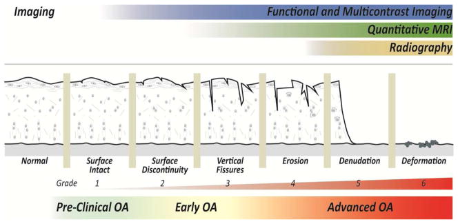
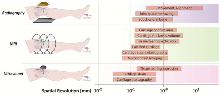
Functional imaging refers broadly to the visualization of organ or tissue physiology using medical image modalities. In load-bearing tissues of the body, including articular cartilage lining the bony ends of joints, changes in strain, stress, and material properties occur in osteoarthritis (OA), providing an opportunity to probe tissue function through the progression of the disease. Here, biomechanical measures in cartilage and related joint tissues are discussed as key imaging biomarkers in the evaluation of OA. Emphasis will be placed on the (1) potential of radiography, ultrasound, and magnetic resonance imaging to assess early tissue pathomechanics in OA, (2) relative utility of kinematic, structural, morphological, and biomechanical measures as functional imaging biomarkers, and (3) improved diagnostic specificity through the combination of multiple imaging biomarkers with unique contrasts, including elastography and quantitative assessments of tissue biochemistry. In comparison to other modalities, magnetic resonance imaging provides an extensive range of functional measures at the tissue level, with conventional and emerging techniques available to potentially to assess the spectrum of preclinical to advance OA.
Keywords: Cartilage degeneration; Elastography; Magnetic resonance imaging (MRI); Radiography; Ultrasound; WOMAC.
Copyright © 2014 Osteoarthritis Research Society International. Published by Elsevier Ltd. All rights reserved.
Conflict of interest statement
There are no conflicts of interest.
Figures
Similar articles
-
Osteoarthritis year in review 2015: imaging.Osteoarthritis Cartilage. 2016 Jan;24(1):49-57. doi: 10.1016/j.joca.2015.07.027. Osteoarthritis Cartilage. 2016. PMID: 26707992 Review.
-
Subchondral bone changes and the impacts on joint pain and articular cartilage degeneration in osteoarthritis.Clin Exp Rheumatol. 2016 Sep-Oct;34(5):929-934. Epub 2016 Aug 31. Clin Exp Rheumatol. 2016. PMID: 27606839 Review.
-
A review on the mechanical quality of articular cartilage - implications for the diagnosis of osteoarthritis.Clin Biomech (Bristol). 2006 Dec;21(10):999-1012. doi: 10.1016/j.clinbiomech.2006.07.001. Epub 2006 Sep 18. Clin Biomech (Bristol). 2006. PMID: 16979270 Review.
-
Chronic in vivo load alteration induces degenerative changes in the rat tibiofemoral joint.Osteoarthritis Cartilage. 2013 Feb;21(2):346-57. doi: 10.1016/j.joca.2012.10.014. Epub 2012 Nov 1. Osteoarthritis Cartilage. 2013. PMID: 23123358 Free PMC article.
-
[Early evaluation of osteoarthritis using objective diagnostic methods].Zhongguo Gu Shang. 2009 May;22(5):402-4. Zhongguo Gu Shang. 2009. PMID: 19522418 Chinese.
Cited by
-
Model for in-vivo estimation of stiffness of tibiofemoral joint using MR imaging and FEM analysis.J Transl Med. 2021 Jul 19;19(1):310. doi: 10.1186/s12967-021-02977-1. J Transl Med. 2021. PMID: 34281578 Free PMC article.
-
Quantitative OCT and MRI biomarkers for the differentiation of cartilage degeneration.Skeletal Radiol. 2016 Apr;45(4):505-16. doi: 10.1007/s00256-016-2334-6. Epub 2016 Jan 19. Skeletal Radiol. 2016. PMID: 26783011
-
Detecting Articular Cartilage and Meniscus Deformation Effects Using Magnetization Transfer Ultrashort Echo Time (MT-UTE) Modeling during Mechanical Load Application: Ex Vivo Feasibility Study.Cartilage. 2021 Dec;13(1_suppl):665S-673S. doi: 10.1177/1947603520976771. Epub 2020 Dec 8. Cartilage. 2021. PMID: 33289401 Free PMC article.
-
Detection of Early-Stage Degeneration in Human Articular Cartilage by Multiparametric MR Imaging Mapping of Tissue Functionality.Sci Rep. 2019 Apr 11;9(1):5895. doi: 10.1038/s41598-019-42543-w. Sci Rep. 2019. PMID: 30976065 Free PMC article.
-
Multiparametric MRI and Computational Modelling in the Assessment of Human Articular Cartilage Properties: A Comprehensive Approach.Biomed Res Int. 2018 May 15;2018:9460456. doi: 10.1155/2018/9460456. eCollection 2018. Biomed Res Int. 2018. PMID: 29862300 Free PMC article.
References
-
- Burstein D, Gray ML, Hartman AL, Gipe R, Foy BD. Diffusion of small solutes in cartilage as measured by nuclear magnetic resonance (NMR) spectroscopy and imaging. Journal of orthopaedic research : official publication of the Orthopaedic Research Society. 1993;11:465–478. - PubMed
Publication types
MeSH terms
Grants and funding
LinkOut - more resources
Full Text Sources
Other Literature Sources
Medical
