. 2014 Nov 7;346(6210):718-722.
doi: 10.1126/science.1258026. Epub 2014 Oct 2.

Structure of the large ribosomal subunit from human mitochondria

Affiliations

Structure of the large ribosomal subunit from human mitochondria

Alan Brown et al. Science. .

Abstract

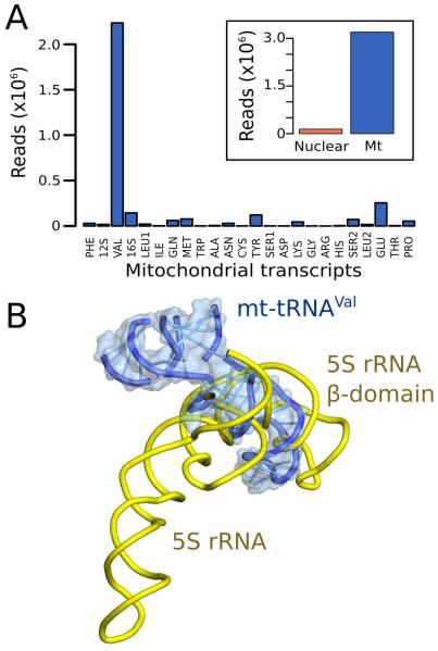
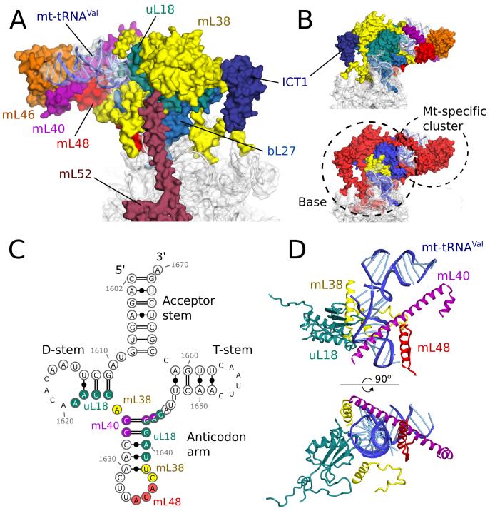
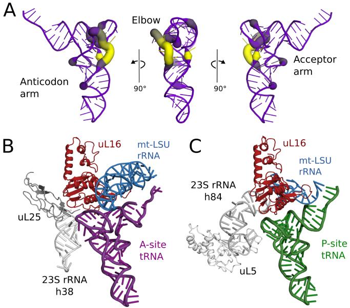
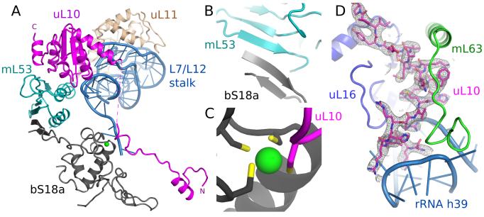
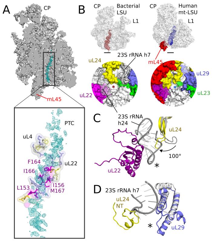
Human mitochondrial ribosomes are highly divergent from all other known ribosomes and are specialized to exclusively translate membrane proteins. They are linked with hereditary mitochondrial diseases and are often the unintended targets of various clinically useful antibiotics. Using single-particle cryogenic electron microscopy, we have determined the structure of its large subunit to 3.4 angstrom resolution, revealing 48 proteins, 21 of which are specific to mitochondria. The structure unveils an adaptation of the exit tunnel for hydrophobic nascent peptides, extensive remodeling of the central protuberance, including recruitment of mitochondrial valine transfer RNA (tRNA(Val)) to play an integral structural role, and changes in the tRNA binding sites related to the unusual characteristics of mitochondrial tRNAs.


Figures

Fig. 1
Fig. 1. Overview of human mt-LSU
(A] Location of proteins in the human mt-LSU, showing (from left to right) solvent-facing, side and exit tunnel views. (B) Views as in A, proteins conserved with bacteria (blue), extensions of homologous proteins (yellow) and mitochondria-specific proteins (red). rRNA is shown in gray.
Fig. 2
Fig. 2. Mt-tRNAVal is part of the human mitoribosome
(A) Mapping of rRNA-sequencing reads to total human (inset) and mitochondrial transcripts. (B) The anticodon stem-loop of mt-tRNAVal binds in a similar position to domain β of 5S rRNA.
Fig. 3
Fig. 3. The central protuberance containing mt-tRNAVal
(A) Relative locations of proteins and mt-tRNAVal in the central protuberance. (B). View of A rotated by 180°, colored by proteins (top) and conservation (bottom) in accordance with Fig. 1. (C) Secondary structure of mt-tRNAVal. Modeled nucleotides are circled, and those interacting with surrounding proteins are colored. (D) The anticodon arm of mt-tRNAVal (blue) interacts extensively with proteins, whereas the acceptor arm is solvent exposed.
Fig. 4
Fig. 4. Co-evolution of mt-tRNAs and their binding sites
(A) Variability in the elbow region of human mt-tRNAs. The deletion of nucleotides relative to a bacterial tRNA (PDB ID: 2WDI) is shown by line color and thickness, with yellow and thick lines indicating most frequently deleted. (B) Modelling a bacterial A-site tRNA (purple) reveals that uL25 and 23S rRNA h38 (both gray) that stabilize the tRNA elbow region are deleted compared to bacterial ribosomes. (C) Similarly, uL5 and 23S rRNA h84 (both gray) that stabilize the elbow region of P-site tRNA (green), are deleted, but elements that bind the anticodon arm are conserved.
Fig. 5
Fig. 5. Remodeling of the L7/L12 stalk
(A) Overview of new elements at the L7/L12 stalk. (B) bS18a forms a shared β-sheet with mL53 to connect the stalk to the body of the mitoribosome. (C) The novel N-terminal extension of uL10 contributes a cysteine residue to a shared zinc-binding motif with bS18a. (D) Density for the N-terminal extension of uL10 that is highly co-ordinated to the body of the ribosome.
Fig. 6
Fig. 6. The exit tunnel
(A) Slice through the mt-LSU showing nascent chain density (cyan) in the exit tunnel. The nascent polypeptide interacts with a β-hairpin of uL22 enriched with hydrophobic residues. (B) The exit tunnel in bacteria (left, red) and human mitoribosomes (right, blue) showing a view of the polypeptide exit site below. The tunnel exit is marked with an asterisk. The polypeptide exit tunnel in mt-LSU is more proteinaceous than in bacteria as a result of two rRNA deletions. (C) Deletion of h7 in bacteria (gray) is compensated by changes to uL29 and an N-terminal extension of uL24. (D) Deletion of h24 (gray) results in the conserved β-hairpin of uL24 rotating closer to the tunnel exit and exposes uL22 to the nascent polypeptide.

References

    1. O’Brien T. Properties of Human Mitochondrial Ribosomes. IUBMB Life. 2003;55:505–513. - PubMed
    1. Liu M, Spremulli L. Interaction of mammalian mitochondrial ribosomes with the inner membrane. J Bio Chem. 2000;275:29400–29406. - PubMed
    1. Skladal D. Minimum birth prevalence of mitochondrial respiratory chain disorders in children. Brain. 2003;126:1905–1912. - PubMed
    1. Boczonadi V, Horvath R. Mitochondria: Impaired mitochondrial translation in human disease. Int J Biochem Cell Biol. 2014;48:77–84. - PMC - PubMed
    1. Sotgia F, et al. Mitochondria “fuel” breast cancer metabolism: fifteen markers of mitochondrial biogenesis label epithelial cancer cells, but are excluded from adjacent stromal cells. Cell cycle. 2012;11:4390–4401. - PMC - PubMed

Publication types

Associated data