Review
. 2014 Oct;27(4):783-822.
doi: 10.1128/CMR.00003-14.

Emerging technologies for the clinical microbiology laboratory

Affiliations
Review

Emerging technologies for the clinical microbiology laboratory

Blake W Buchan et al. Clin Microbiol Rev. 2014 Oct.

Abstract

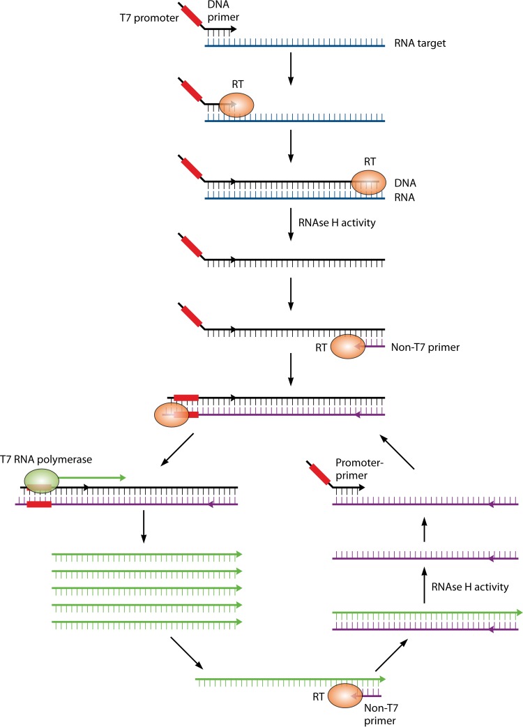
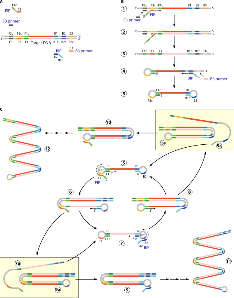
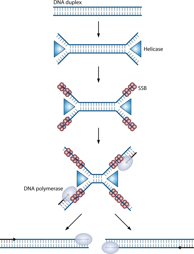
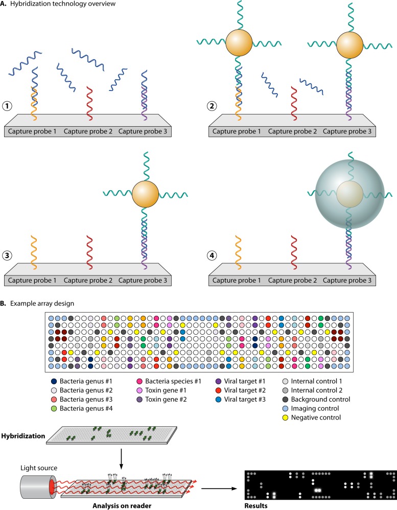
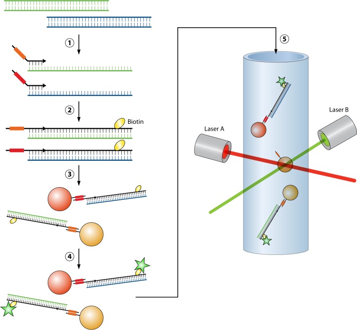
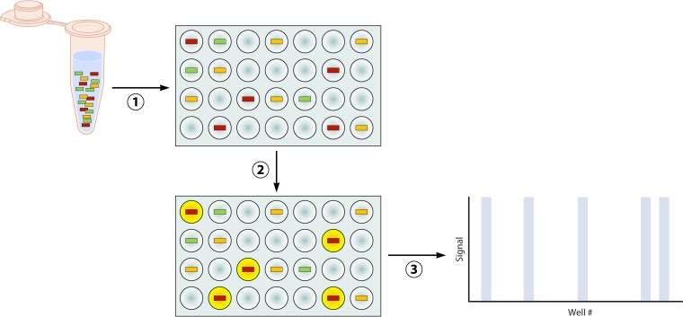
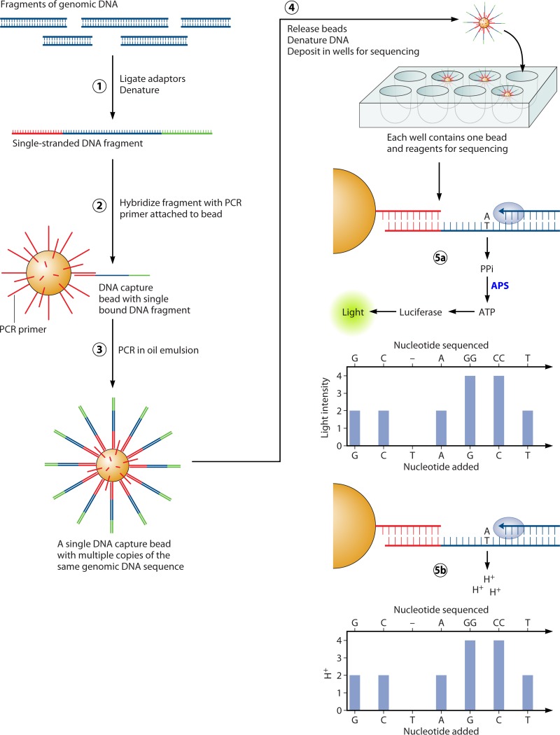
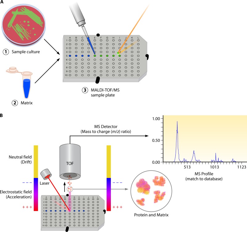
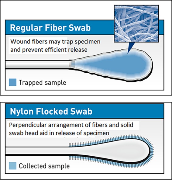
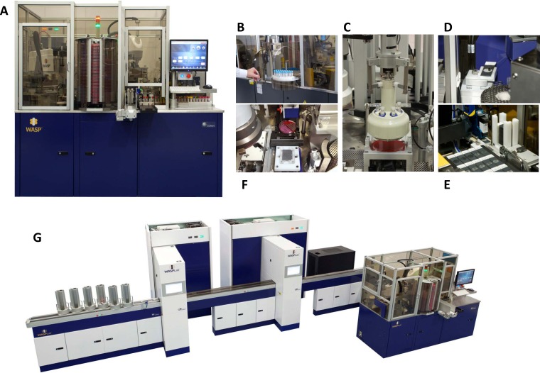
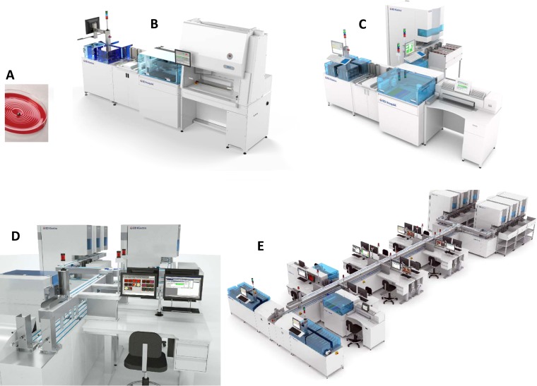
In this review we examine the literature related to emerging technologies that will help to reshape the clinical microbiology laboratory. These topics include nucleic acid amplification tests such as isothermal and point-of-care molecular diagnostics, multiplexed panels for syndromic diagnosis, digital PCR, next-generation sequencing, and automation of molecular tests. We also review matrix-assisted laser desorption ionization-time of flight (MALDI-TOF) and electrospray ionization (ESI) mass spectrometry methods and their role in identification of microorganisms. Lastly, we review the shift to liquid-based microbiology and the integration of partial and full laboratory automation that are beginning to impact the clinical microbiology laboratory.


Figures

FIG 1
FIG 1
Transcription-mediated amplification (TMA). The single-stranded RNA target is bound by a cDNA primer engineered to contain a T7 viral RNA polymerase promoter sequence (red box). Reverse transcriptase (RT) extends the DNA primer to form an RNA-cDNA duplex, and the RNA template strand is degraded by RNase H activity. A second primer anneals to the single-stranded cDNA (black) and is extended by RT, which incorporates the T7 promoter into the double-stranded DNA sequence. T7 RNA polymerase recognizes the incorporated T7 promoter sequence and synthesizes 100 to 1,000 copies of single-strand RNA amplicon (green). These amplicons serve both as a target for detection probes and as a single-stranded template for subsequent rounds of amplification using the non-T7 primer to initiate cDNA synthesis by RT.
FIG 2
FIG 2
Loop-mediated isothermal amplification (LAMP). (A) LAMP-based amplification requires 4 primers complementary to 6 different regions of the nucleic acid target (F1, F2, F3, B1, B2, and B3). The “inner primers” FIP and BIP each contain two regions complementary to the target sequence; one anneals to the template strand (F2 and B2), and one anneals to the complementary strand (F1c and B1c). The “outer primers” (F3 and B3) are complementary to a single sequence upstream of FIP and BIP, respectively. (B) Replication initiates with annealing and extension of the FIP and BIP “inner primers.” The “outer primers” F3 and B3 anneal upstream of FIP and BIP and are extended, which displaces the strands initiated by the FIP and BIP inner primers. The displaced strands form 5′ loop structures through complementary binding, resulting in a single-strand “dumbbell” structure. (C) The single-strand “dumbbell” serves as the template for subsequent rounds of amplification using the FIP and BIP primers to initiate elongation. Single-stranded template is maintained through formation of loop structures which can be extended to displace newly synthesized double-strand product (C5 through C8). (Adapted from reference with permission from Macmillan Publishers Ltd.)
FIG 3
FIG 3
Helicase-dependent amplification (HDA). HDA uses the UvrD (helicase) (blue triangles) and MutL (accessory protein required for efficient UvrD loading to DNA) enzymes from E. coli to catalyze temperature-independent creation of a single-stranded DNA template for nucleic acid amplification. The UvrD/MutL complex unwinds double-stranded DNA to form an open complex. Single-strand binding proteins (SSB) (red circles) bind to the denatured strands to prevent association of the complementary strands. Specific primers are designed to anneal to the target sequence, and DNA polymerase (gray oval) extends the primers to the generate target amplicon. This amplicon serves as the template for subsequent rounds of amplification. (Adapted from reference with permission [copyright Wiley-VCH Verlag GmbH & Co. KGaA].)
FIG 4
FIG 4
Verigene solid-phase microarray. (A) Single-stranded, target-specific capture probes are arrayed spatially and immobilized onto the surface of a glass slide. The nucleic acid target (PCR amplicon or extracted nucleic acid) is denatured and applied to the glass slide. If present, the target nucleic acid will anneal to the complementary capture probe. Gold microspheres coated with single-stranded nucleic acid complementary to a different region of the target sequence are added and anneal to the capture probe-target sequence hybrid to form a “sandwich” nucleic acid structure. The array is washed to remove unbound nucleic acid and gold microparticles. Application of colloidal silver increases the size of the bound microspheres to increase the sensitivity of detection. (B) Target-specific capture probes, along with internal controls, are spotted in triplicate to different locations on the glass slide to ensure consistency of the annealing and hybridization steps and increase accuracy of results. Target detection is accomplished using a light source shown across the plane of the array. If present, bound silver microspheres diffract the light, which is then detected by an optical camera in the array reader.
FIG 5
FIG 5
xTAG liquid-phase microarray. Target sequences (blue and green) are amplified using multiplex PCR. Following amplification, a second set of target-specific primers containing “universal tag sequences” (orange and red boxes) unique to each target primer are used for a primer extension reaction. During primer extension, a biotin label is also incorporated into the amplicon. Labeled amplicons are then incubated with polystyrene microbeads. Microbeads are uniquely colored, allowing differentiation of up to 100 different types of microbeads by the analyzer. Each color bead is also coated with a single-strand nucleic acid probe complementary to one of the universal tag sequences (antitag). Amplicons labeled with universal tag sequences will hybridize to the microbeads containing the antitag. Additionally, a streptavidin-fluorophore conjugate (green star) is added and hybridizes to biotin-labeled amplicons immobilized on the beads. Following hybridization steps, beads are analyzed using a cell sorter equipped with two lasers. The first detects the presence of the fluorophore conjugated to biotin, indicating the presence of an amplicon bound to a specific microbead. The second laser interrogates the bead to determine which dye is present, thereby identifying the specific target amplicon present. The center bead in step 5 lacks amplicon and thus would be negative for the biotin-fluorophore signal. This bead would not be analyzed by the second laser.
FIG 6
FIG 6
Digital PCR. A nucleic acid template containing target sequences (colored boxes) in the original sample is diluted into individual microwells (plate PCR, pictured) or picoliter droplets (emulsion PCR) such that each well or droplet contains one or zero copies of the target sequence. Following partitioning of the specimen, endpoint PCR is carried out and amplicon is detected using fluorescent dyes or probes. Each well will be either positive or negative for fluorescent signal depending on the presence of the target sequence and resulting amplicon (yellow circles correspond to blue bars on the graph). The number of wells or droplets positive for fluorescent signal (yellow circles) directly corresponds to the number of specific target sequences (red boxes) present in the original sample.
FIG 7
FIG 7
Next generation sequencing by synthesis. Next generation sequencing by 454 (pyrosequencing) and Ion Torrent (semiconductor sequencing) utilize similar techniques to generate sequence information. In an initial step, genomic or amplified DNA to be sequenced is fragmented and single-strand overhangs are enzymatically removed. Synthetic nucleic acid adaptors (red and green) are ligated to each end of the target nucleic acid fragment. The modified target is then denatured and incubated with microbeads coated with a single-stranded capture probe (red) complementary to one of the adaptors (red). Hybridization immobilizes the target onto the surface of the bead, and beads are then partitioned into oil emulsion droplets containing reagents required for PCR. The PCR amplifies the target sequence, resulting in a single bead coated with thousands of identical copies of the target sequence. Following PCR, the beads are partitioned into microwells for sequencing. Each well contains the reagents required for sequencing with the exception of nucleotides. For both pyrosequencing and semiconductor sequencing, wells are washed with each of the four nucleoside bases in sequential order. 5a, in pyrosequencing, addition of a complementary nucleoside results in the release of pyrophosphate (PPi). The PPi is converted to ATP by ATP sulfurylase in the presence of adenosine 5′ phosphosulfate (APS), which is used to drive light production by luciferase. 5b, in semiconductor sequencing, release of H+ upon addition of a complementary nucleoside results in a change in pH, which is measured by a semiconductor in the bottom on the sequencing well. In both cases, the intensity of the signal (light or pH change) is proportional to the number of nucleotides incorporated. Therefore, addition of two consecutive nucleotides (e.g., GG) will generate a signal approximately twice the intensity of that generated by a single nucleoside insertion.
FIG 8
FIG 8
Matrix-assisted laser desorption ionization–time of flight mass spectrometry (MALDI-TOF MS). (A) Preparation of samples for analysis by MALDI-TOF MS can be either by direct transfer of a bacterial colony to the sample plate using a sterile implement (green spots) or as a liquid supernatant following an extraction procedure (blue spots). In either method, the analyte is allowed to dry before being overlaid with a weak acid matrix material. (B) Analysis of the analyte begins with exposure to a laser, which ionizes and desorbs analyte form the sample plate. The created ions are accelerated through the time of flight vacuum tube by application of an electrostatic field until they reach the MS detector. Ions with a larger mass-to-charge ratio (m/z) will take longer to traverse the time of flight tube than ions with a smaller m/z. An MS profile is created with the m/z of each ion species plotted on the x axis and the relative abundance of each m/z ion species on the y axis. This MS profile is compared to a reference spectral library of defined spectra to establish a “best-match” identification of the isolate being analyzed. (Reprinted from reference with permission.)
FIG 9
FIG 9
Comparison of wound fiber swabs to flocked swabs. Traditional swabs are constructed by winding fiber strands around the tip of a straight shaft to create a wound fiber bulb for collection of the specimen. Winding of fibers creates a “net” which may entrap microorganisms and prevent efficient release onto solid or liquid culture medium. Flocked swabs are composed of a solid bulbous core at the tip of the swab which is coated with perpendicular fibers. This arrangement allows for more efficient release of microorganisms collected in a specimen onto culture medium.
FIG 10
FIG 10
Walk-away specimen processor (WASP). The multifunctional WASP core unit (A) includes two independently operating robotic arms capable of decapping, recapping, and inoculating up to 180 solid agar plates per hour. The core unit is also equipped with a vortex for sample mixing and a “tool belt” which can accommodate three different-size reusable calibrated inoculating loops and a blunt tipped colony-picking instrument which can be accessed by the robotic arm as needed. Culture media are housed in a 9-silo carousel which can accommodate up to 350 standard agar plates. Liquid media for broth culture are housed in a “warehouse carousel” located on the reverse side of the WASP (B). The WASP is also equipped with a barcode label reader capable of reading specimen barcodes and a printer which automatically prints and applies labels to all corresponding plates prior to inoculation. The core WASP unit can be equipped with optional disk dispenser (C) for application of antibiotic disks to inoculated media, a Gram slide prep module for automated preparation of slides (D and E), and a stage for automated transfer of isolated colonies and matrix material to a MALDI target plate for analysis using MALDI-TOF MS (F). Total laboratory automation features the WASP core unit connected via conveyer tracks to smart incubators equipped with high-resolution cameras for imaging of culture plates (G). (Courtesy of Copan Diagnostics.)
FIG 11
FIG 11
BD Kiestra total laboratory automation (TLA). The BD Kiestra TLA system is composed of task-specific modules. The stand-alone InoqulA module is capable of automated plate inoculation and streaking of up to 400 plates per hour using the roll bead method (A). Fully automated inoculation, streaking, barcoding, and sorting of inoculated media can be conducted by a combination of the SorterA, BarcodeA, and InoqulA modules, an optional biosafety cabinet for manual plating of nonliquid specimens can also be integrated into front-end workflow (B). Work Cell Automation (WCA) (C) incorporates an ErgonomicA technologist workstation (D) and incubators equipped with high-resolution cameras for imaging culture plates (ReadA). Work Cell Automation modules can be configured into large or small total laboratory automation systems (E) with multiple InoqulA, ErgonomicA, and ReadA modules to accommodate additional specimen volume in medium- to high-throughput microbiology laboratories. (Courtesy of BD Kiestra.)
None
None

Similar articles

Cited by

References

    1. Hodinka RL. 2013. Point: is the era of viral culture over in the clinical microbiology laboratory? J. Clin. Microbiol. 51:2–4. 10.1128/JCM.02593-12 - DOI - PMC - PubMed
    1. Clark AE, Kaleta EJ, Arora A, Wolk DM. 2013. Matrix-assisted laser desorption ionization-time of flight mass spectrometry: a fundamental shift in the routine practice of clinical microbiology. Clin. Microbiol. Rev. 26:547–603. 10.1128/CMR.00072-12 - DOI - PMC - PubMed
    1. Burd EM. 2010. Validation of laboratory-developed molecular assays for infectious diseases. Clin. Microbiol. Rev. 23:550–576. 10.1128/CMR.00074-09 - DOI - PMC - PubMed
    1. Valenstein PN. 1990. Evaluating diagnostic tests with imperfect standards. Am. J. Clin. Pathol. 93:252–258 - PubMed
    1. CLSI. 2005. User varification of performance for precisioin and trueness. Approved guideline, 2nd ed. CLSI document EP15-A2. CLSI, Wayne, PA

Publication types