Review
. 2014 Oct;27(4):870-926.
doi: 10.1128/CMR.00109-13.

Coagulase-negative staphylococci

Affiliations
Review

Coagulase-negative staphylococci

Karsten Becker et al. Clin Microbiol Rev. 2014 Oct.

Abstract

The definition of the heterogeneous group of coagulase-negative staphylococci (CoNS) is still based on diagnostic procedures that fulfill the clinical need to differentiate between Staphylococcus aureus and those staphylococci classified historically as being less or nonpathogenic. Due to patient- and procedure-related changes, CoNS now represent one of the major nosocomial pathogens, with S. epidermidis and S. haemolyticus being the most significant species. They account substantially for foreign body-related infections and infections in preterm newborns. While S. saprophyticus has been associated with acute urethritis, S. lugdunensis has a unique status, in some aspects resembling S. aureus in causing infectious endocarditis. In addition to CoNS found as food-associated saprophytes, many other CoNS species colonize the skin and mucous membranes of humans and animals and are less frequently involved in clinically manifested infections. This blurred gradation in terms of pathogenicity is reflected by species- and strain-specific virulence factors and the development of different host-defending strategies. Clearly, CoNS possess fewer virulence properties than S. aureus, with a respectively different disease spectrum. In this regard, host susceptibility is much more important. Therapeutically, CoNS are challenging due to the large proportion of methicillin-resistant strains and increasing numbers of isolates with less susceptibility to glycopeptides.


Figures

FIG 1
FIG 1
Time line of the discovery of the species belonging to the genus Staphylococcus. Coagulase-negative species are shown in blue; coagulase-positive and coagulase-variable species are shown in red (note that only S. schleiferi subsp. coagulans is coagulase positive). Note that at the times of establishment of the first three species designations, S. aureus, S. epidermidis, and S. saprophyticus, these terms comprised a broader content than that accepted today. In particular, S. epidermidis and S. saprophyticus were used to describe nonpathogenic, saprophytic staphylococci (and other Gram-positive cocci occurring in clusters).
FIG 2
FIG 2
Clinical and epidemiological schema of staphylococcal species, based on the categorization of coagulase as a major virulence factor and its resulting impact on human health.
FIG 3
FIG 3
Phylogenetic separation of staphylococcal species and subspecies (ssp.), extended by key diagnostic characteristics as proposed by Lamers et al. (32).
FIG 4
FIG 4
Pathogenesis of catheter-related infections and factors influencing biofilm genesis. The image shows the three-step process of biofilm formation on the surface of an intravascular catheter, with rapid initial adhesion and attachment of CoNS microorganisms to the polymer foreign body surface resulting in a monolayer (1), followed by a prolonged accumulation phase which involves cell proliferation, intercellular adhesion processes, and maturation (2 and 3). (4) Finally, microorganisms may disaggregate from the macrocolony and drift into the bloodstream, resulting in metastatic and embolic complications.
FIG 5
FIG 5
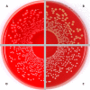
Sections of Columbia blood agar plates showing grayish, hemolytic colonies of S. haemolyticus (the color of this photograph was modified to enhance visibility of the weak hemolysis zones [arrows] surrounding the colonies, resulting in a nonnatural reddish tinge) (A); orange, nonhemolytic colonies of S. chromogenes (B); creamy, nonhemolytic colonies of S. lugdunensis (C); and whitish, nonhemolytic colonies of S. saprophyticus subsp. saprophyticus (D).
FIG 6
FIG 6
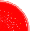
Columbia blood agar plate showing an isogenic S. epidermidis strain pair displaying both the normal (arrows) and SCV (dashed arrows) phenotypes. The normal phenotype (NP) on this plate was the result of a spontaneous reversion of the SCV back to the NP.
FIG 7
FIG 7
Highly simplified schema (not true to scale) of the composition of the SCC family integrated into the S. aureus chromosome (blue). Some examples of various SCC types, including those organized in composite islands, are given. Basic structures comprise the mec gene complex (red), the ccr gene complex (yellow), and the joining regions (gray). Some SCCs additionally contain (i) resistance operons acting against antibiotic agents (violet), such as fusidic acid (fusC); (ii) resistance operons for metalloids and transition metals with toxic/bactericidal properties (green), such as arsenic (ars), cadmium (cad), copper (copB and copC), and mercury (mer); (iii) virulence genes (brown), such as the capsule polysaccharide gene (cap1); (iv) other genes (orange), such as the genes composing the arginine catabolic mobile element (arc), the copper-translocating P-type ATPase gene (copA), and the potassium-transporting ATPase genes (kdp); (v) further penicillin-binding protein 4 (PBP4) genes (pink), such as pbp4; and (vi) pseudogenes.
None
None
None

References

    1. Kloos WE, Bannerman TL. 1994. Update on clinical significance of coagulase-negative staphylococci. Clin. Microbiol. Rev. 7:117–140 - PMC - PubMed
    1. Pfaller MA, Herwaldt LA. 1988. Laboratory, clinical, and epidemiological aspects of coagulase-negative staphylococci. Clin. Microbiol. Rev. 1:281–299 - PMC - PubMed
    1. Taponen S, Pyörälä S. 2009. Coagulase-negative staphylococci as cause of bovine mastitis—not so different from Staphylococcus aureus? Vet. Microbiol. 134:29–36. 10.1016/j.vetmic.2008.09.011 - DOI - PubMed
    1. Irlinger F. 2008. Safety assessment of dairy microorganisms: coagulase-negative staphylococci. Int. J. Food Microbiol. 126:302–310. 10.1016/j.ijfoodmicro.2007.08.016 - DOI - PubMed
    1. Billroth T. 1874. Untersuchungen über die Vegetationsformen von Coccobacteria septica und den Antheil, welchen sie an der Entstehung und Verbreitung der accidentellen Wundkrankheiten haben: Versuch einer wissenschaftlichen Kritik der verschiedenen Methoden antiseptischer Wundbehandlung. G Reimer, Berlin, Germany

Publication types

MeSH terms