Review
. 2014 Oct;27(4):980-1024.
doi: 10.1128/CMR.00126-13.

Cryptococcus gattii infections

Affiliations
Review

Cryptococcus gattii infections

Sharon C-A Chen et al. Clin Microbiol Rev. 2014 Oct.

Abstract

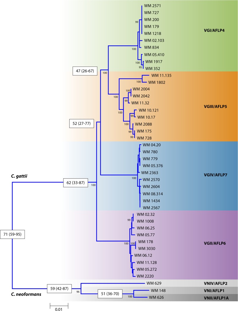
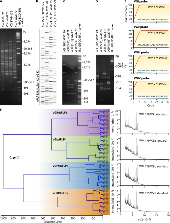
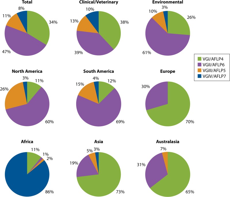
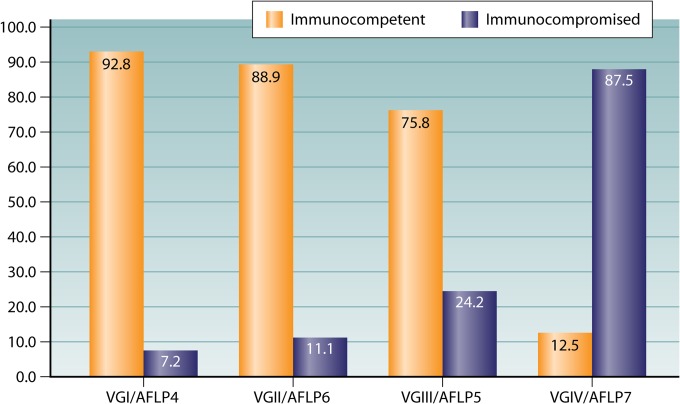
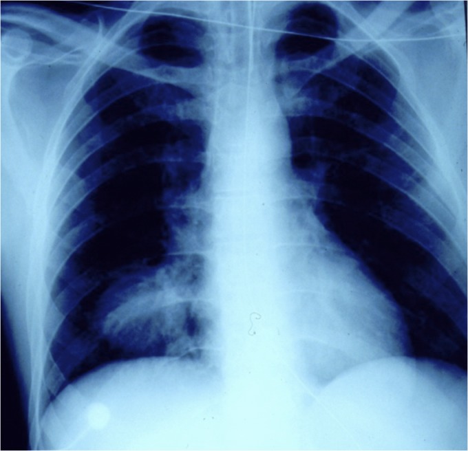
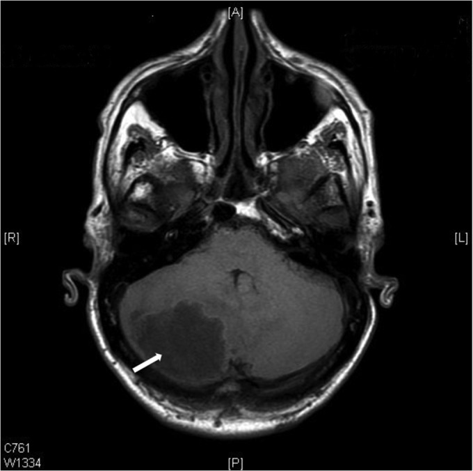
Understanding of the taxonomy and phylogeny of Cryptococcus gattii has been advanced by modern molecular techniques. C. gattii probably diverged from Cryptococcus neoformans between 16 million and 160 million years ago, depending on the dating methods applied, and maintains diversity by recombining in nature. South America is the likely source of the virulent C. gattii VGII molecular types that have emerged in North America. C. gattii shares major virulence determinants with C. neoformans, although genomic and transcriptomic studies revealed that despite similar genomes, the VGIIa and VGIIb subtypes employ very different transcriptional circuits and manifest differences in virulence phenotypes. Preliminary evidence suggests that C. gattii VGII causes severe lung disease and death without dissemination, whereas C. neoformans disseminates readily to the central nervous system (CNS) and causes death from meningoencephalitis. Overall, currently available data indicate that the C. gattii VGI, VGII, and VGIII molecular types more commonly affect nonimmunocompromised hosts, in contrast to VGIV. New, rapid, cheap diagnostic tests and imaging modalities are assisting early diagnosis and enabling better outcomes of cerebral cryptococcosis. Complications of CNS infection include increased intracranial pressure, severe neurological sequelae, and development of immune reconstitution syndrome, although the mortality rate is low. C. gattii VGII isolates may exhibit higher fluconazole MICs than other genotypes. Optimal therapeutic regimens are yet to be determined; in most cases, initial therapy with amphotericin B and 5-flucytosine is recommended.


Figures

FIG 1
FIG 1
Rooted neighbor-joining tree inferred from the concatenated MLST typing scheme sequences (CAP59, GPD1, LAC1, SOD1, URA5, and PLB1 genes and IGS) of 10 strains of each major C. gattii molecular type rooted with the standard strains of the three major C. neoformans molecular types. Numbers on branches are bootstrap support values obtained from 1,000 pseudoreplicates using the program MEGA version 5.05. Numbers in the white boxes indicate estimated diversion times based on the 10 genetic concatenated loci (ACT1, LAC1, IDE1, ITS1/2, IGS, PLB1, RPB1, RPB2, TEF1, and URA5), generated with the program BEAST.
FIG 2
FIG 2
Identification of the major molecular types within C. gattii. (A) PCR fingerprinting generated with the primer M13; (B) AFLP profiles generated with the 6-carboxyfluorescein (FAM) label AC+G kit; (C) PLB1 gene RFLP profiles identified via digestion with AvaI; (D) URA5 gene RFLP profiles identified via double digestion with Sau96I and HhaI; (E) PLB1 gene hyperbranched rolling-circle amplification; (F) MALDI-TOF MS profiles obtained from the reference strains of each major molecular type.
FIG 3
FIG 3
Distribution of the four major molecular types of C. gattii identified among a total of 980 clinical/veterinary and environmental global isolates. Shown is the distribution of the four major molecular types of C. gattii, combining clinical/veterinary (n = 615) and environmental (n = 365) isolates (top) from North America (including Mexico) (n = 162), South America (n = 367), and Europe (n = 44) (middle) and from Africa (n = 64), Asia (n = 78), and Australasia (n = 265) (bottom).
FIG 4
FIG 4
Global distribution of the four major molecular types of C. gattii, based on 980 isolates.
FIG 5
FIG 5
Percentages of 256 clinical isolates obtained from immunocompetent and immunocompromised patients (HIV positive) and from patients with other risk factors per major molecular type, identified by URA5 RFLP analysis, PCR fingerprinting, or AFLP analysis, for which clinical data were available. Other risk factors are alcoholism, corticosteroid use, disorder T immunity, diabetes, leukemia, systemic lupus erythematosus, transplant, and tumor.
FIG 6
FIG 6
Chest X ray of a patient with a large mass lesion in the right lower lobe. Bronchoscopy and collection of bronchoalveolar lavage fluid showed multiple encapsulated yeasts upon cytological examination, and a culture grew Cryptococcus gattii. (Reprinted from reference with permission of the publisher. Copyright 2013 UpToDate, Inc. [www.uptodate.com].)
FIG 7
FIG 7
Computerized tomographic scan of brain showing a large mass lesion in the left cerebellar hemisphere (indicated by arrow). A provisional diagnosis of a cerebral neoplasm was made. Excision biopsy of the lesion showed encapsulated yeasts, and Cryptococcus gattii was isolated in culture.
None
None
None

References

    1. Fraser JA, Giles SS, Wenink EC, Geunes-Boyer SG, Wright JR, Diezmann S, Allen A, Stajich JE, Dietrich FS, Perfect JR, Heitman J. 2005. Same-sex mating and the origin of the Vancouver Island Cryptococcus gattii outbreak. Nature 437:1360–1364. 10.1038/nature04220 - DOI - PubMed
    1. Kwon-Chung KJ, Bennett JE, Theodore TS. 1978. Cryptococcus bacillisporus sp. nov.: serotype B-C of Cryptococcus neoformans. Int. J. Syst. Bacteriol. 28:616–620. 10.1099/00207713-28-4-616 - DOI
    1. Findley K, Sun S, Fraser JA, Hsueh YP, Averette AF, Li W, Dietrich FS, Heitman J. 2012. Discovery of a modified tetrapolar sexual cycle in Cryptococcus amylolentus and the evolution of MAT in the Cryptococcus species complex. PLoS Genet. 8:e1002528. 10.1371/journal.pgen.1002528 - DOI - PMC - PubMed
    1. Fell JW, Boekhout T, Fonseca A, Scorzetti G, Statzell-Tallman A. 2000. Biodiversity and systematics of basidiomycetous yeasts as determined by large-subunit rDNA D1/D2 domain sequence analysis. Int. J. Syst. Evol. Microbiol. 50(Part 3):1351–1371. 10.1099/00207713-50-3-1351 - DOI - PubMed
    1. Sanfelice F. 1895. Ueber einen neuen pathogenen Blastomyceten welcher innerhalb der Gewebe unter Bildung kalkartig aussehender Massen degeneriert. Zentralbl. Bakteriol. Parasit. Infect. Hyg. 18:521–526

Publication types

MeSH terms

LinkOut - more resources