

18-Year-Old Woman with an Embryonal Rhabdomyosarcoma of the Uterus in Statu Nascendi
- PMID: 25278623
- PMCID: PMC4168320
- DOI: 10.1055/s-0032-1328076
18-Year-Old Woman with an Embryonal Rhabdomyosarcoma of the Uterus in Statu Nascendi
Abstract
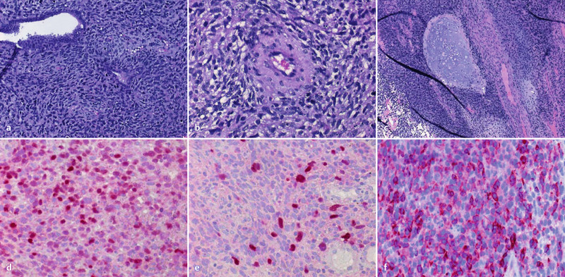
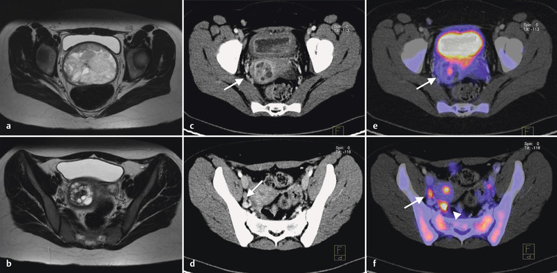
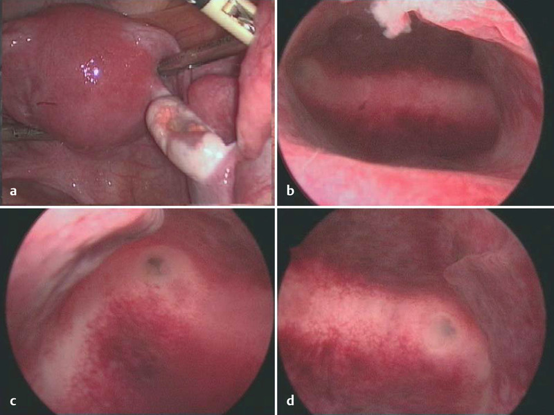
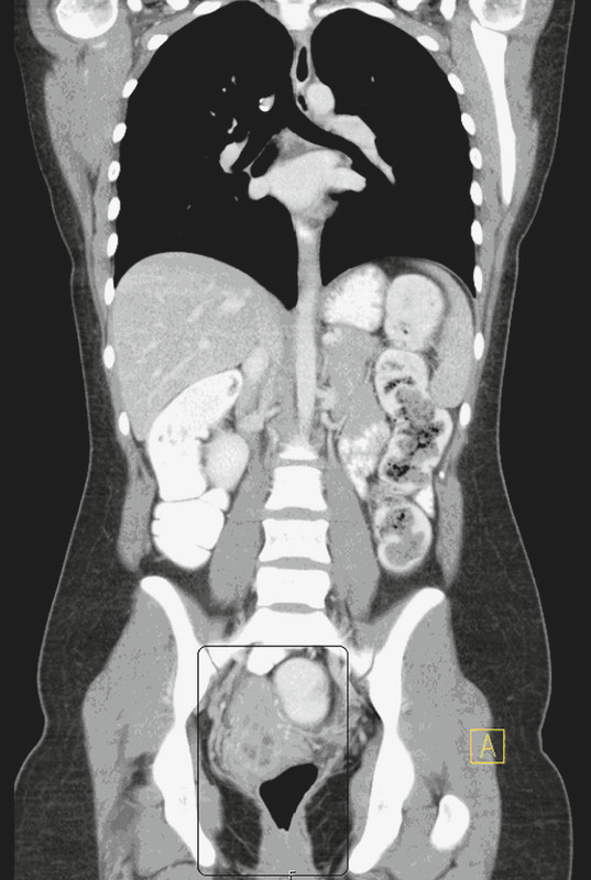
Background: We report a case of an 18-year-old woman with an embryonal rhabdomyosarcoma in statu nascendi. Case: A fist-sized embryonal rhabdomyosarcoma of the uterus filling the vaginal vault was diagnosed in an adolescent with virgo intacta suffering from therapy resistant vaginal discharge, bleeding and bulging mass for six months. Further imaging revealed one suspicious pelvine lymph node. Excision of the tumour including the intracervical stalk was performed and followed by systemic multiagent chemotherapy. PET-CT scan presented a complete response after the third cycle. Histological complete response was shown by laparoscopic dissection of regional pelvic lymph nodes and curettage. Conclusion: Uterine Rhabdomyosarcoma should be considered as differential diagnosis of therapy resistant vaginal flour and bleeding in young women. Fertility-sparing therapy is possible in selected exceptional cases.
Hintergrund: Wir berichten über eine 18-jährige Patientin mit Erstdiagnose eines embryonalen Rhabdomyosarkoms des Uterus in statu nascendi. Fallbericht: Bei einer 18-jährigen Patientin G0, virgo intacta fiel ein übelriechender, zerfallender Tumor im Bereich der Scheide auf. Die durchgeführte Biopsie zeigte einen malignen mesenchymalen Tumor mit Verdacht auf embryonales Rhabdomyosarkom. Bei starker vaginaler Blutung wurde am Aufnahmetag ein MRT des Beckens durchgeführt. Hier wurde ebenfalls der Verdacht auf ein Leiomyosarkom ausgehend von der Vagina mit Zervixbeteiligung und zusätzlich eines metastasensuspekten Lymphknoten rechts pelvin diagnostiziert. Nach vaginaler Partialexzision des Primarius wurde mit systemischer Chemotherapie begonnen. Die Reevaluation mittels PET-CT nach dem 3. Zyklus der systemischen Therapie zeigte eine ycT0-ycN0-cM0-Situation. Die Komplettremission konnte durch laparoskopisch durchgeführte pelvine Lymphnodektomie rechts und fraktionierte Kürettage histologisch bestätigt werden. Fazit: Bei den jungen Patientinnen mit therapieresistenten vaginalen Fluor und Blutungen sollte bei der Differenzialdiagnose an ein uterines Rhabdomyosarkom gedacht werden. In Ausnahmefällen kann die Tumortherapie auch fertilitätserhaltend durchgeführt werden.
Keywords: embryonal rhabdomyosarcoma; sarcoma botryoides; uterine sarcoma; uterine tumor.
Conflict of interest statement
Figures
References
-
- Ferguson S E, Gerald W, Barakat R R. et al. Clinicopathologic features of rhabdomyosarcoma of gynecologic origin in adults. Am J Surg Pathol. 2007;31:382–389. - PubMed
-
- da Silva B B, Dos Santos A R, Bosco Parentes-Vieira J. et al. Embryonal rhabdomyosarcoma of the uterus associated with uterine inversion in an adolescent: a case report and published work review. J Obstet Gynaecol Res. 2008;34(4 Pt 2):735–738. - PubMed
-
- Perrone T, Carson L F, Dehner L P. Rhabdomyosarcoma with heterologous cartilage of the uterine cervixa clinicopathologic and immunohistochemical study of an aggressive neoplasm in a young female. Med Pediatr Oncol. 1990;18:72–76. - PubMed
-
- Copeland L J, Sneige N, Stringer A. et al. Alveolar rhabdomyosarcoma of the female genitalia. Cancer. 1985;56:849–855. - PubMed
-
- Montag T WD, Ablaing G, Schlaerth J B. et al. Embryonal rhabdomyosarcoma of the uterine corpus and cervix. Gynecol Oncol. 1986;25:171–194. - PubMed
LinkOut - more resources
Full Text Sources