Review
. 2014 Sep 18:8:290.
doi: 10.3389/fncel.2014.00290. eCollection 2014.

A synaptic mechanism for network synchrony

Affiliations
Review

A synaptic mechanism for network synchrony

Simon T Alford et al. Front Cell Neurosci. .

Abstract

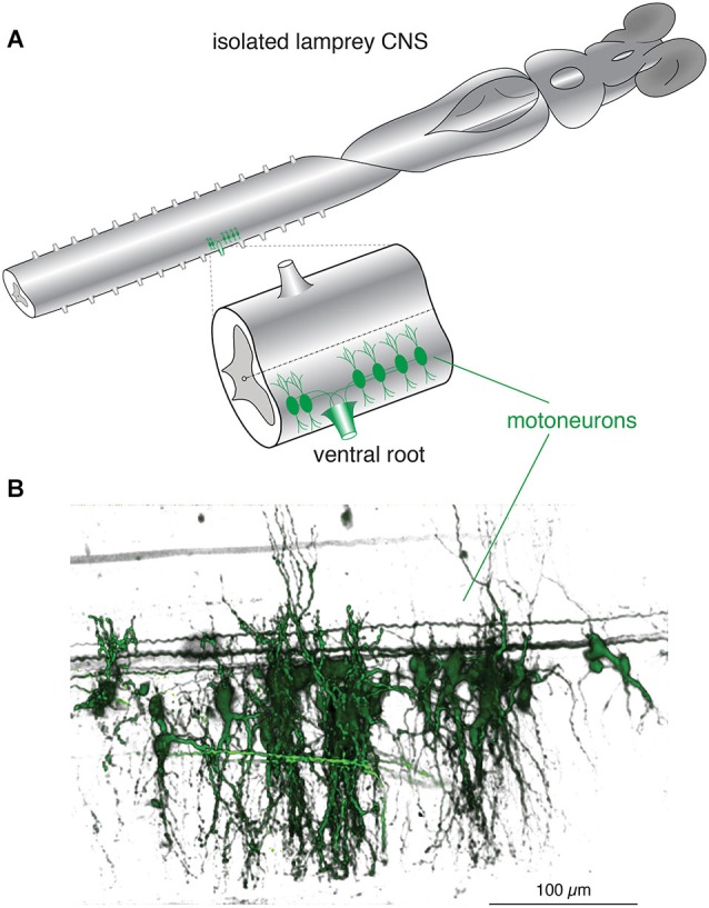
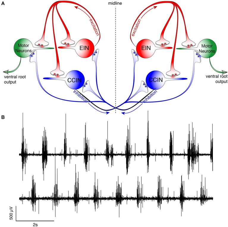
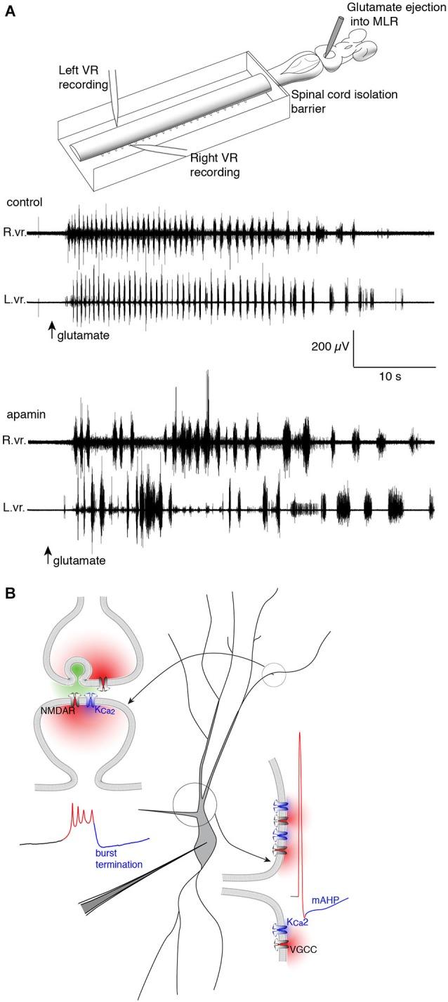
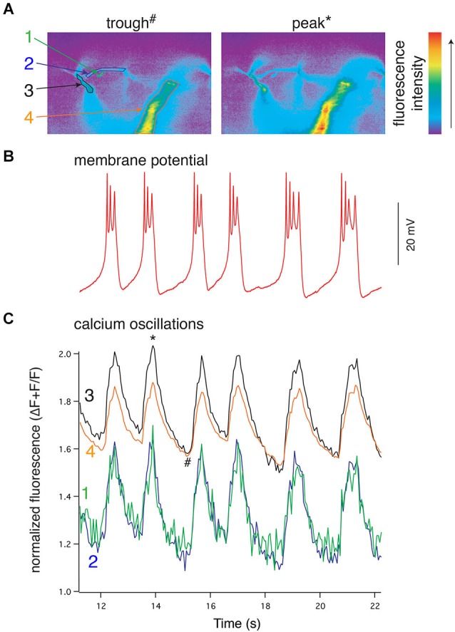
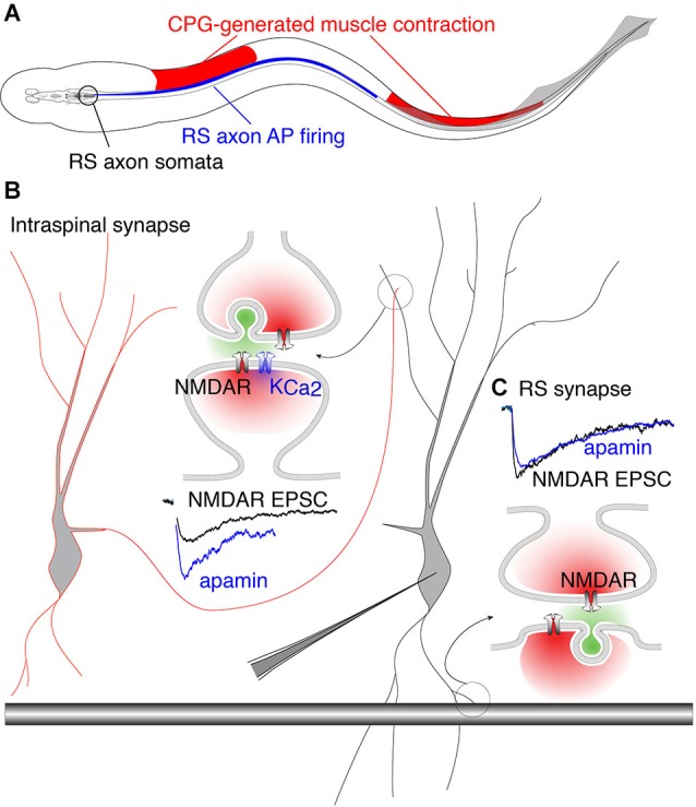
Within neural networks, synchronization of activity is dependent upon the synaptic connectivity of embedded microcircuits and the intrinsic membrane properties of their constituent neurons. Synaptic integration, dendritic Ca(2+) signaling, and non-linear interactions are crucial cellular attributes that dictate single neuron computation, but their roles promoting synchrony and the generation of network oscillations are not well understood, especially within the context of a defined behavior. In this regard, the lamprey spinal central pattern generator (CPG) stands out as a well-characterized, conserved vertebrate model of a neural network (Smith et al., 2013a), which produces synchronized oscillations in which neural elements from the systems to cellular level that control rhythmic locomotion have been determined. We review the current evidence for the synaptic basis of oscillation generation with a particular emphasis on the linkage between synaptic communication and its cellular coupling to membrane processes that control oscillatory behavior of neurons within the locomotor network. We seek to relate dendritic function found in many vertebrate systems to the accessible lamprey central nervous system in which the relationship between neural network activity and behavior is well understood. This enables us to address how Ca(2+) signaling in spinal neuron dendrites orchestrate oscillations that drive network behavior.

Keywords: KCa2; NMDA; SK2; calcium; dendrites; lamprey; locomotion; oscillation.


Figures

Figure 1
Figure 1
Lamprey spinal motoneurons have a complex dendritic architecture. (A) Schematic representation of an isolated lamprey brain and spinal cord. Spinal motoneurons and their complete dendritic architecture can be retrogradely labeled through an intramuscular injection of a dextran-conjugated fluorescent dye. Labeling (green) is visible on the side and segment of injection through axons converging into ventral roots (VRs) and to their respective neurons. Expansion shows a single spinal segment with multiple motoneurons labeled as in (B). (B) A 3D reconstruction of motoneurons labeled from one spinal ventral root to emphasize the complexity of their structure and dendritic trees. Neurons were labeled by injecting the muscle wall of an animal with 5 μL of 5 mM Oregon Green 488 BAPTA1 Dextran. After 24 h the animal was sacrificed and the spinal cord fixed and cleared. A confocal stack was imaged to generate the full extent of the motoneurons in one spinal segment (Viana di Prisco and Alford, 2004).
Figure 2
Figure 2
Schematic representation of the lamprey spinal central pattern generator. (A) Spinal CPG neurons receive both ipsilateral glutamatergic (red) input from excitatory interneurons (EINs, red) and contralaterally projecting glycinergic inhibition (blue) from reciprocally inhibiting, crossed glycinergic interneurons (CCINs, blue). Output of the CPG occurs from motoneurons (green), which directly synapse onto myotomal cells of the trunk musculature to cause muscle contraction producing rhythmic locomotion. (B) Output pattern recorded using glass suction electrodes from paired, contralateral (top vs. bottom traces) VRs showing alternating bursting of the spinal network during rhythmic locomotion. The reciprocally connected network described in (A) prevents excitation of the contralateral spinal cord when the ipsilateral side is bursting for each cycle (burst-to-burst), leading each side of the spinal cord to be precisely 180° out-of-phase from the other (Alford et al., 2003).
Figure 3
Figure 3
Repolarizing KCa2 channels are spatially segregated in lamprey spinal VHNs according to their function and mechanism of activation. (A) Top: An isolated lamprey CNS can be used to study the brain and spinal circuits controlling locomotion. Pressure-ejection of L-glutamate into the lamprey mesencephalic locomotor region (MLR) induces short episodes of fictive locomotion, the electrophysiological correlate of locomotion. Using a dual-pool recording chamber, pharmacological agents can be selectively applied to the spinal cord, without interfering with descending commands originating in the brainstem that initiate and maintain locomotion. Locomotor bursts are recorded directly from left and right VRs. Bottom: A long locomotor episode with regular, alternating bursts (control) follows after a puff of glutamate into the MLR (arrow, glutamate). Blockade of KCa2 channels with the selective antagonist, apamin, decreases the burst frequency and disrupts the alternating locomotor rhythm. This demonstrates the necessity of KCa2 channels for correct alternation and regularity of the locomotor rhythm (Nanou et al., 2013). (B) The effect of KCa2 channel blockade on locomotion can be explained by the role the channel plays at the cellular level. Within VHNs, KCa2 currents may be evoked either at synapses (top left) whereby synaptic release of glutamate activates NMDAR-mediated Ca2+ entry and thereby closely located KCa2 channels. It is this KCa2-mediated current that is critical for the termination of NMDA-TTX oscillations (blue portion of trace) shown below recorded from somatic microelectrode recordings. KCa2-mediated currents are also responsible for the mAHP seen following action potential firing shown at bottom left. However, this current is activated following Ca2+ entry from VGCCs.
Figure 4
Figure 4
NMDA-evoked, TTX-resistant oscillations in lamprey VHNs show simultaneous oscillations in Ca2+ throughout the dendritic tree. (A) VHN neurons were labeled with the Ca2+-sensitive dye, Oregon Green 488 BAPTA1, by pressure injection from a recording microelectrode and recorded during oscillations evoked by application of NMDA (100 μM) in TTX (1 μM). Pseudocolored, raw images are shown from the trough of the hyperpolarization (left, denoted by # in (C)) and the peak of depolarization (right, denoted by * in (C)). Colored numbers and arrows point to discrete regions of interest whose fluorescence measurements are shown in (C). Fluorescence intensity scale shown to the right. (B) Current clamp recording of the membrane potential oscillations. (C) Simultaneous to the membrane potential oscillations in (B), Ca2+ recorded using the fluorescent dye Oregon Green 488 BAPTA1 shows transient increases in concentration in the dendrites. In the proximal dendrites, the oscillations are above a higher baseline Ca2+ evoked by NMDA application than that recorded in the distal dendrites, however, all recorded regions of the dendrites exhibit these Ca2+ oscillations. All Ca2+ fluorescence is normalized to the fluorescence at rest prior to the application of NMDA. The regions recorded are indicated by colored numbers in (A) and (C) (Alford et al., 2003).
Figure 5
Figure 5
Phase-matched and phase-mismatched excitatory synapses in the spinal cord of lamprey. (A) As the lamprey swims it generates a traveling wave from head to tail. The sinusoidal body curvature illustrated here represents a single moment in body movement during a bout of swimming. During swimming, command excitation is continually provided by RS axons whose somata in the brainstem (encircled in black) fire (blue) in phase with the rostral spinal CPG neurons. This is illustrated by the rostro-caudal overlap of red and blue. It is the output of spinal segments that causes ipsilateral muscle contraction (red). Due to the speed at which the action potential (AP) propagates along RS axons, the AP invades more caudal areas of the spinal cord whose associated muscles do not undergo contraction because the CPG wave (responsible for contraction) travels at a delay relative to the AP. This leads to regions along the spinal cord where AP firing overlaps with inhibited musculature (illustrated by overlap of blue and white regions in the middle). This would predictably lead VHN excitation at inappropriate times during the swim cycle. This phase mismatch between RS axons and CPG neurons may be avoided by synapse-specific KCa2 channel activation. (B) Circuit model in which excitation from EINs (red outlined cell) projects to other VHNs (black outlined cell) locally within the spinal cord. NMDAR currents from these neurons (black trace, NMDAR EPSC) are enhanced by the addition of apamin, the specific KCa2 channel antagonist, to block KCa2 currents (blue trace). (C) RS synapses from large descending axons (black shaded bar) which project throughout the spinal cord, show NMDAR currents (black trace, NMDAR EPSC) that are unaffected by apamin (blue trace) (Alpert and Alford, 2013).

Similar articles

Cited by

References

    1. Abalo X. M., Villar-Cheda B., Meléndez-Ferro M., Pérez-Costas E., Anadón R., Rodicio M. C. (2007). Development of the serotonergic system in the central nervous system of the sea lamprey. J. Chem. Neuroanat. 34, 29–46 10.1016/j.jchemneu.2007.03.010 - DOI - PubMed
    1. Akita T., Kuba K. (2000). Functional triads consisting of ryanodine receptors, Ca(2+) channels and Ca(2+)-activated K(+) channels in bullfrog sympathetic neurons. Plastic modulation of action potential. J. Gen. Physiol. 116, 697–720 10.1085/jgp.116.5.697 - DOI - PMC - PubMed
    1. Alford S., Frenguelli B. G., Schofield J. G., Collingridge G. L. (1993). Characterization of Ca2+ signals induced in hippocampal CA1 neurones by the synaptic activation of NMDA receptors. J. Physiol. 469, 693–716 - PMC - PubMed
    1. Alford S., Grillner S. (1990). CNQX and DNQX block non-NMDA synaptic transmission but not NMDA-evoked locomotion in lamprey spinal cord. Brain Res. 506, 297–302 10.1016/0006-8993(90)91266-j - DOI - PubMed
    1. Alford S., Schwartz E., Viana di Prisco G. (2003). The pharmacology of vertebrate spinal central pattern generators. Neuroscientist 9, 217–228 10.1177/1073858403009003014 - DOI - PubMed

LinkOut - more resources