

Neural precursor cells in the ischemic brain - integration, cellular crosstalk, and consequences for stroke recovery
- PMID: 25278840
- PMCID: PMC4165213
- DOI: 10.3389/fncel.2014.00291
Neural precursor cells in the ischemic brain - integration, cellular crosstalk, and consequences for stroke recovery
Abstract
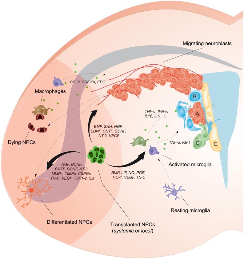
After an ischemic stroke, neural precursor cells (NPCs) proliferate within major germinal niches of the brain. Endogenous NPCs subsequently migrate toward the ischemic lesion where they promote tissue remodeling and neural repair. Unfortunately, this restorative process is generally insufficient and thus unable to support a full recovery of lost neurological functions. Supported by solid experimental and preclinical data, the transplantation of exogenous NPCs has emerged as a potential tool for stroke treatment. Transplanted NPCs are thought to act mainly via trophic and immune modulatory effects, thereby complementing the restorative responses initially executed by the endogenous NPC population. Recent studies have attempted to elucidate how the therapeutic properties of transplanted NPCs vary depending on the route of transplantation. Systemic NPC delivery leads to potent immune modulatory actions, which prevent secondary neuronal degeneration, reduces glial scar formation, diminishes oxidative stress and stabilizes blood-brain barrier integrity. On the contrary, local stem cell delivery allows for the accumulation of large numbers of transplanted NPCs in the brain, thus achieving high levels of locally available tissue trophic factors, which may better induce a strong endogenous NPC proliferative response. Herein we describe the diverse capabilities of exogenous (systemically vs. locally transplanted) NPCs in enhancing the endogenous neurogenic response after stroke, and how the route of transplantation may affect migration, survival, bystander effects and integration of the cellular graft. It is the authors' claim that understanding these aspects will be of pivotal importance in discerning how transplanted NPCs exert their therapeutic effects in stroke.
Keywords: blood–brain barrier; brain plasticity; cell therapy; neurogenesis; neuroprotection; stroke.
Figures
Similar articles
-
Transduction of neural precursor cells with TAT-heat shock protein 70 chaperone: therapeutic potential against ischemic stroke after intrastriatal and systemic transplantation.Stem Cells. 2012 Jun;30(6):1297-310. doi: 10.1002/stem.1098. Stem Cells. 2012. PMID: 22593021
-
Effects of acute versus post-acute systemic delivery of neural progenitor cells on neurological recovery and brain remodeling after focal cerebral ischemia in mice.Cell Death Dis. 2014 Aug 21;5(8):e1386. doi: 10.1038/cddis.2014.359. Cell Death Dis. 2014. PMID: 25144721 Free PMC article.
-
Transplantation of iPS cell-derived neural progenitors overexpressing SDF-1α increases regeneration and functional recovery after ischemic stroke.Oncotarget. 2017 Oct 31;8(57):97537-97553. doi: 10.18632/oncotarget.22180. eCollection 2017 Nov 14. Oncotarget. 2017. PMID: 29228630 Free PMC article.
-
Stem Cell Therapy in Brain Trauma: Implications for Repair and Regeneration of Injured Brain in Experimental TBI Models.In: Kobeissy FH, editor. Brain Neurotrauma: Molecular, Neuropsychological, and Rehabilitation Aspects. Boca Raton (FL): CRC Press/Taylor & Francis; 2015. Chapter 42. In: Kobeissy FH, editor. Brain Neurotrauma: Molecular, Neuropsychological, and Rehabilitation Aspects. Boca Raton (FL): CRC Press/Taylor & Francis; 2015. Chapter 42. PMID: 26269908 Free Books & Documents. Review.
-
Brain regeneration in physiology and pathology: the immune signature driving therapeutic plasticity of neural stem cells.Physiol Rev. 2011 Oct;91(4):1281-304. doi: 10.1152/physrev.00032.2010. Physiol Rev. 2011. PMID: 22013212 Free PMC article. Review.
Cited by
-
Neural Transplants From Human Induced Pluripotent Stem Cells Rescue the Pathology and Behavioral Defects in a Rodent Model of Huntington's Disease.Front Neurosci. 2020 Sep 18;14:558204. doi: 10.3389/fnins.2020.558204. eCollection 2020. Front Neurosci. 2020. PMID: 33071737 Free PMC article.
-
Systemic Factors Trigger Vasculature Cells to Drive Notch Signaling and Neurogenesis in Neural Stem Cells in the Adult Brain.Stem Cells. 2019 Mar;37(3):395-406. doi: 10.1002/stem.2947. Epub 2018 Dec 30. Stem Cells. 2019. PMID: 30431198 Free PMC article.
-
Actin Alpha 2 Downregulation Inhibits Neural Stem Cell Proliferation and Differentiation into Neurons through Canonical Wnt/β-Catenin Signaling Pathway.Oxid Med Cell Longev. 2022 Feb 9;2022:7486726. doi: 10.1155/2022/7486726. eCollection 2022. Oxid Med Cell Longev. 2022. PMID: 35186189 Free PMC article.
-
Neural stem cell transplantation in ischemic stroke: A role for preconditioning and cellular engineering.J Cereb Blood Flow Metab. 2017 Jul;37(7):2314-2319. doi: 10.1177/0271678X17700432. Epub 2017 Mar 17. J Cereb Blood Flow Metab. 2017. PMID: 28303738 Free PMC article. Review.
-
Harnessing migraines for neural regeneration.Neural Regen Res. 2018 Apr;13(4):609-615. doi: 10.4103/1673-5374.230275. Neural Regen Res. 2018. PMID: 29722303 Free PMC article. Review.
References
Publication types
Grants and funding
LinkOut - more resources
Full Text Sources
Other Literature Sources