. 2012 Mar;6(1):49-52.
doi: 10.5704/MOJ.1203.003.

Intraosseous synovial sarcoma of the proximal femur: Case report

Affiliations

Intraosseous synovial sarcoma of the proximal femur: Case report

M Zulkarnaen et al. Malays Orthop J. 2012 Mar.

Abstract

Abstract: Synovial sarcoma is primarily a soft tissue malignancy that most often affects adolescents and young adults. It very rarely presents as a primary bone tumour and has only been reported in nine other cases to date. We report a case of primary synovial sarcoma arising from the proximal femur in a 57-year-old man.

Key words: Synovial sarcoma, primary bone tumour.


Figures
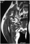
<b>Fig. 1</b>: Magnetic resonance imaging (MRI) showing an
intraosseous lesion (arrow) at the proximal femur with an intertrochanteric fracture.
Fig. 1: Magnetic resonance imaging (MRI) showing an intraosseous lesion (arrow) at the proximal femur with an intertrochanteric fracture.
<b>Fig. 2a</b>: Gross specimen of the proximal femur with a normal looking muscle cuff surrounding the tumour and fracture site (red arrow).
The distal femur shaft was cut at 3 cm distal to the tumour margin based on pre-chemotherapy MRI measurement.
Fig. 2a: Gross specimen of the proximal femur with a normal looking muscle cuff surrounding the tumour and fracture site (red arrow). The distal femur shaft was cut at 3 cm distal to the tumour margin based on pre-chemotherapy MRI measurement.
<b>Fig. 2b</b>: Cut section of the proximal femur showing the fleshy grey, haemorrhagic tumour (red arrow) involving the metaphysis and diaphysis. Note that there is minimal involvement of the surrounding soft tissue.
Fig. 2b: Cut section of the proximal femur showing the fleshy grey, haemorrhagic tumour (red arrow) involving the metaphysis and diaphysis. Note that there is minimal involvement of the surrounding soft tissue.
<b>Fig. 3</b>: Histological image showing glandular formations (red arrow) which is positive for pan-Cytokeratin, Epithelial Membrane Antigen (EMA) and Cytokeratin 7 (CK 7) antibodies. Other areas show predominantly spindled
malignant cells.
Fig. 3: Histological image showing glandular formations (red arrow) which is positive for pan-Cytokeratin, Epithelial Membrane Antigen (EMA) and Cytokeratin 7 (CK 7) antibodies. Other areas show predominantly spindled malignant cells.

References

    1. Weiss WS, Goldblum JR, Enzinger FM. Enzinger and Weiss' soft tissue tumors.5th ed. Philadelphia: Mosby Elsevier; 2008.
    1. Fletcher CDM, Unni KK, Mertens F. World Health Organization., International Agency for Research on Cancer. Pathology and genetics of tumours of soft tissue and bone. Lyon: IARC Press; 2002.
    1. Cohen IJ, Issakov J, Avigad S, Stark B, Meller I, Zaizov R. Synovial sarcoma of bone delineated by spectral karyotyping. Lancet. 1997;350(9092):1679–1680. - PubMed
    1. NG Beck S, Raskin KA, Schwab JH. Intraosseous Synovial Sarcoma of the Proximal Tibia. International Journal of Surgical Oncology. 2001;2001:1–5. - PMC - PubMed
    1. Hiraga H, Nojima T, Isu K, Yamashiro K, Yamawaki S, Nagashima K. Histological and molecular evidence of synovial sarcoma of bone. A case report. J Bone Joint Surg [AM] 1999;81(4):558–563. - PubMed

LinkOut - more resources