

Peripapillary and macular choroidal thickness in glaucoma
- PMID: 25279115
- PMCID: PMC4181196
Peripapillary and macular choroidal thickness in glaucoma
Abstract
Purpose: To compare choroidal thickness (CT) between individuals with and without glaucomatous damage and to explore the association of peripapillary and submacular CT with glaucoma severity using spectral domain optical coherence tomography (SD-OCT).
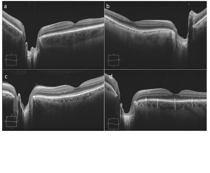
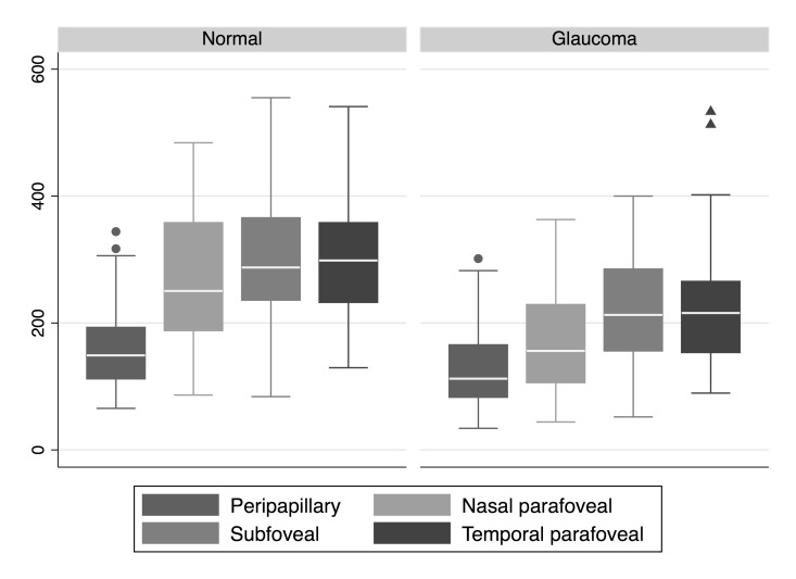
Methods: Ninety-one eyes of 20 normal subjects and 43 glaucoma patients from the UCLA SD-OCT Imaging Study were enrolled. Imaging was performed using Cirrus HD-OCT. Choroidal thickness was measured at four predetermined points in the macular and peripapillary regions, and compared between glaucoma and control groups before and after adjusting for potential confounding variables.
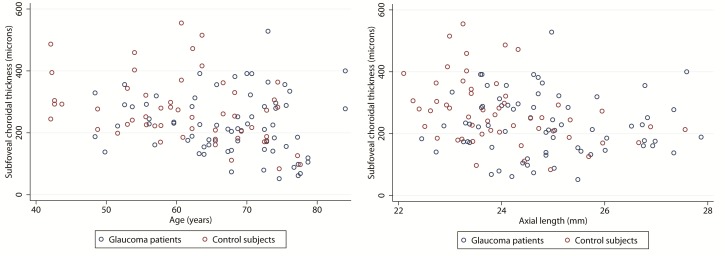
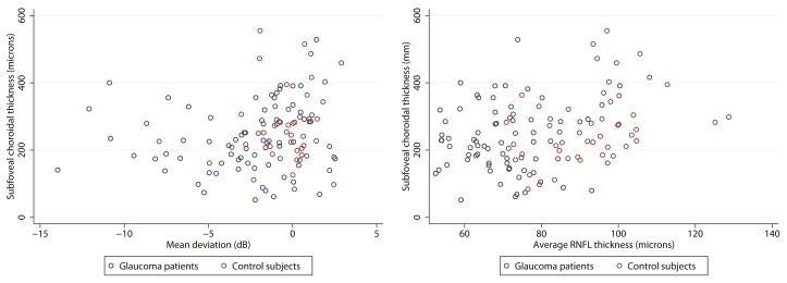
Results: The average (± standard deviation) mean deviation (MD) on visual fields was -0.3 (±2.0) dB in controls and -3.5 (±3.5) dB in glaucoma patients. Age, axial length and their interaction were the most significant factors affecting CT on multivariate analysis. Adjusted average CT (corrected for age, axial length, their interaction, gender and lens status) however, was not different between glaucoma patients and the control group (P=0.083) except in the temporal parafoveal region (P=0.037); nor was choroidal thickness related to glaucoma severity (r=-0.187, P=0.176 for correlation with MD, r=-0.151, P=0.275 for correlation with average nerve fiber layer thickness).
Conclusions: Choroidal thickness of the macular and peripapillary regions is not decreased in glaucoma. Anatomical measurements with SD-OCT do not support the possible influence of the choroid on the pathophysiology of glaucoma.
Keywords: Choroidal Thickness; Glaucoma; Macula; Peripapillary; Spectral-Domain Optical Coherence Tomography.
Figures
References
-
- Schuman JS, Hee MR, Arya AV, Pedut-Kloizman T, Puliafito CA, Fujimoto JG, et al. Optical coherence tomography: a new tool for glaucoma diagnosis. Curr Opin Ophthalmol. 1995;6:89–95. - PubMed
-
- Lee KY, Tomidokoro A, Sakata R, Konno S, Mayama C, Saito H, et al. Cross-sectional anatomic configurations of peripapillary atrophy evaluated with spectral domain-optical coherence tomography. Invest Ophthalmol Vis Sci. 2010;51:666–671. - PubMed
-
- Deokule S, Vizzeri G, Boehm AG, Bowd C, Medeiros FA, Weinreb RN. Correlation among choroidal, parapapillary, and retrobulbar vascular parameters in glaucoma. Am J Ophthalmol. 2009;147:736–743. - PubMed
-
- Leske MC, Heijl A, Hyman L, Bengtsson B, Dong LM, Yang Z. Predictors of long-term progression in the early manifest glaucoma trial. Ophthalmology. 2007;114:1965–1972. - PubMed
-
- Leske MC, Wu SY, Hennis A, Honkanen R, Nemesure B. Risk factors for incident open-angle glaucoma: The Barbados Eye Studies. Ophthalmology. 2008;115:85–93. - PubMed
LinkOut - more resources
Full Text Sources
Miscellaneous