

Bladder extragastrointestinal stromal tumor in an adolescent patient: A case-based review
- PMID: 25279181
- PMCID: PMC4179831
- DOI: 10.3892/mco.2014.366
Bladder extragastrointestinal stromal tumor in an adolescent patient: A case-based review
Abstract
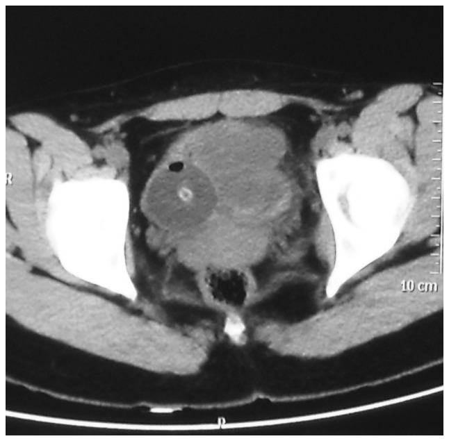
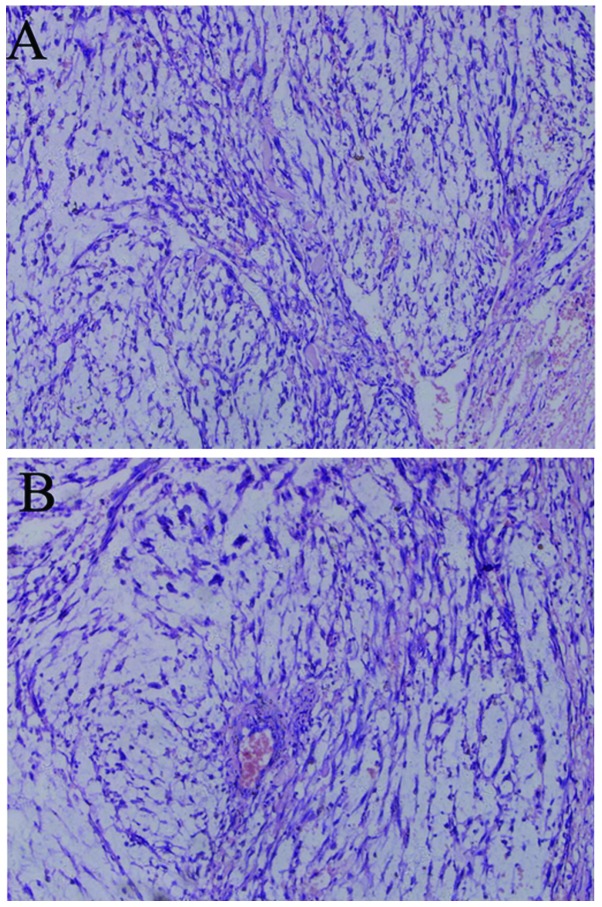
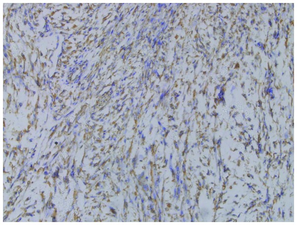
Extragastrointestinal stromal tumors (EGISTs) are mesenchymal tumors occurring outside the gastrointestinal tract, with histological, immunohistochemical and molecular genetic characteristics similar to those of gastrointestinal stromal tumors (GISTs). The immunohistochemical examination usually demonstrates a positive expression for CD117. GISTs are rare neoplasms and EGISTs are even less common. This is the report of a case of EGIST in the urinary bladder of a 15-year-old adolescent female patient who presented with painless gross hematuria. Pelvic computed tomography revealed an irregular soft tissue density mass, sized 5.7×4.8 cm, in the bladder. Partial cystectomy was performed in April, 2011. There was no recurrence during follow-up over the next 35 months, as determined by transabdoninal ultrasonography and cystoscopic examination. The patient in this study did not receive any molecular-targeted drugs. To the best of our knowledge, this is the first reported case of an EGIST of the urinary bladder in an adolescent patient.
Keywords: CD117; adolescent; bladder; extragastrointestinal stromal tumor.
Figures
References
-
- Llenas-Garcia J, Guerra-Vales JM, Moreno A, Ibarrola C, Castelbon FJ, Fernandez-Ruiz M, Meneu JC, Ballestin C, Moreno E. Primary extragastrointestinal stromal tumors in the omentum and mesentery: a clinicopathological and immunohistochemical study. Hepatogastroenterology. 2008;55:1002–1005. - PubMed
-
- Miettinen M, Monihan JM, Sarlomo-Rikala M, Kovatich AJ, Carr NJ, Emory TS, Sobin LH. Gastrointestinal stromal tumors/smooth muscle tumors (GISTs) primary in the omentum and mesentery: clinicopathologic and immunohistochemical study of 26 cases. Am J Surg Pathol. 1999;23:1109–1118. - PubMed
-
- Reith JD, Goldblum JR, Lyles RH, Weiss SW. Extragastrointestinal (soft tissue) stromal tumors: an analysis of 48 cases with emphasis on histologic predictors of outcome. Mod Pathol. 2000;13:577–585. - PubMed
-
- Fletcher CD, Berman JJ, Corless C, et al. Diagnosis of gastrointestinal stromal tumors: A consensus approach. Hum Pathol. 2002;33:459–465. - PubMed
-
- Lasota J, Carlson JA, Miettinen M. Spindle cell tumor of urinary bladder serosa with phenotypic and genotypic features of gastrointestinal stromal tumor. Arch Pathol Lab Med. 2000;124:894–897. - PubMed
LinkOut - more resources
Full Text Sources
Other Literature Sources
Miscellaneous