. 2014 Oct 3;9(10):e108904.
doi: 10.1371/journal.pone.0108904. eCollection 2014.

Different patterns of punctate white matter lesions in serially scanned preterm infants

Affiliations

Different patterns of punctate white matter lesions in serially scanned preterm infants

Karina J Kersbergen et al. PLoS One. .

Erratum in

  • PLoS One. 2014;9(11):e114704

Abstract

Background and purpose: With the increased use of MRI in preterm infants, punctate white matter lesions (PWML) are more often recognized. The aim of this study was to describe the incidence and characteristics of these lesions as well as short-term outcome in a cohort of serially scanned preterm infants, using both conventional imaging, diffusion (DWI) and susceptibility (SWI) weighted imaging.

Materials and methods: 112 preterm infants with 2 MRIs in the neonatal period, with evidence of punctate white matter lesions, were included. Appearance, lesion load, location, and abnormalities on DWI and SWI were scored and outcome data were collected.

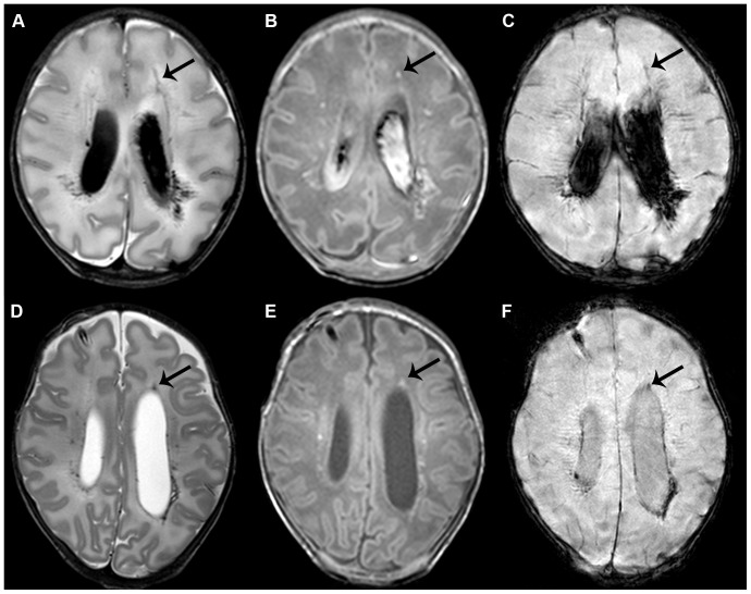
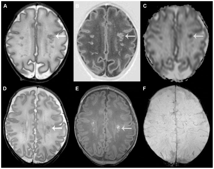
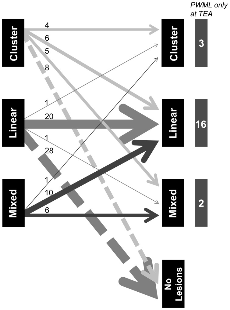
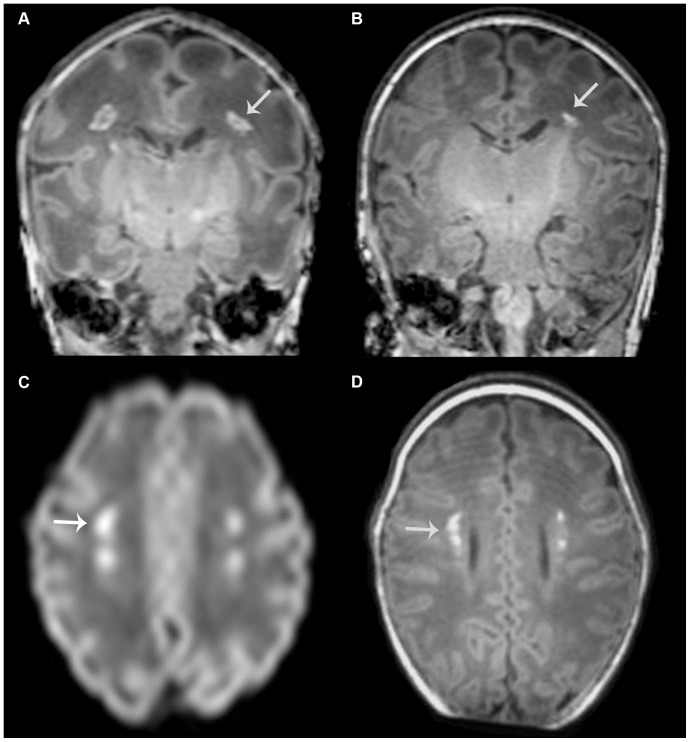
Results: Different patterns of punctate white matter lesions did appear: a linear appearance associated with signal loss on SWI, and a cluster appearance associated with restricted diffusion on DWI on the first MRI. Cluster and mixed lesions on the first scan changed in appearance in over 50% on the second scan, whereas linear lesions generally kept their appearance. Lesions were only visible on the early scan in 33%, and were only seen at term equivalent age in 20%. Nine infants developed cerebral palsy, due to additional overt white matter lesions in six.

Conclusion: Two patterns of punctate white matter lesions were identified: one with loss of signal on SWI in a linear appearance, and the other with DWI lesions with restricted diffusion in a cluster appearance. These different patterns are suggestive of a difference in underlying pathophysiology. To reliably classify PWML in the preterm infant in either pattern, an early MRI with DWI and SWI sequences is required.


Conflict of interest statement

Competing Interests: The authors have declared that no competing interests exist.

Figures

Figure 1
Figure 1. Punctate white matter lesions with a linear pattern.
Early (A–C) and term equivalent (D–F) scans in an infant of 32 weeks gestation with lesions in a mixed, though mainly linear pattern. T2-weighted imaging shows bilateral lesions in the white matter adjacent to the ventricles (A). Lesions are also visible on T1-weighted imaging, additionally showing a small subdural hemorrhage (B). The arrow indicates one of the lesions, visible on all images. Also note the grade III intraventricular hemorrhage. SWI shows signal loss in the areas of the lesions, suggestive of a hemorrhagic origin (C). Additionally, some small frontal lesions can be identified, that do not show up on SWI, and show a cluster appearance. At term equivalent age, lesion load has greatly diminished and the intraventricular hemorrhage has largely resolved on T2- and T1-weighted imaging (D, E). A subcutaneous reservoir has been inserted to treat post-hemorrhagic ventricular dilatation. SWI still shows signal loss with blood residue being most clearly visible on this sequence (F). Outcome was favorable with a cognitive composite score of 115 and a total motor composite score of 124 on the Bayley scales at two years corrected age.
Figure 2
Figure 2. Punctate white matter lesions with a cluster pattern.
Early (A–C) and term equivalent (D–F) scans in an infant of 31+3 weeks gestation with lesions in a cluster pattern. T2-weighted imaging shows multiple lesions throughout the white matter (A) that are also clearly visible on inversion recovery imaging (B). The apparent diffusion coefficient -map shows restricted diffusion at the site of the lesions (C). The SWI sequence (not shown) did not show any signal loss. The arrow indicates one of the lesions, visible on all images. At term equivalent age, lesion load has diminished. Lesions are now better appreciated on T1-weighted imaging (E), compared with T2-weighted imaging (D). Again, SWI does not show signal loss (F). Outcome was favorable with a developmental quotient of 94 on the Griffiths scales at 18 months corrected age.
Figure 3
Figure 3. Changes in PWML appearance over time.
This figure depicts the changes in appearance of PWML between both scans. Early imaging is represented on the left and term equivalent imaging on the right. The number of infants per category is depicted by the thickness of the arrow and also printed above each arrow. Especially cluster and mixed lesions will change appearance, whereas linear lesions remain linear in the majority of the infants, or are no longer visible at TEA. At the right side of the figure, infants are depicted that only showed PWML at TEA.
Figure 4
Figure 4. PWML in an infant without additional lesions who developed cerebral palsy.
T1-weighted images at early (born at 31 weeks, postmenstrual age at scan 33 weeks, (A)) and term equivalent age (B) in an infant with lesions in a cluster pattern. Note that the lesions seem to be directly in the path of the corticospinal tracts (see arrow for an example). Lesions show high signal intensity, suggestive of restricted diffusion, on DWI of the early scan (C) and these are visible in the same location on T1-weighted imaging at term equivalent age (D). No signal intensity changes were seen on SWI on either scan and no additional lesions were identified. At term equivalent age, the posterior limb of the internal capsule appears to be less well developed on the right side. This infant subsequently developed a mild asymmetric bilateral spastic cerebral palsy, with her left leg being most severely affected.

References

    1. Debillon T, N'Guyen S, Muet A, Quere MP, Moussaly F, et al. (2003) Limitations of ultrasonography for diagnosing white matter damage in preterm infants. Arch Dis Child Fetal Neonatal Ed 88: F275–F279. - PMC - PubMed
    1. Maalouf EF, Duggan PJ, Counsell SJ, Rutherford MA, Cowan F, et al. (2001) Comparison of findings on cranial ultrasound and magnetic resonance imaging in preterm infants. Pediatrics 107: 719–727. - PubMed
    1. Miller SP, Cozzio CC, Goldstein RB, Ferriero DM, Partridge JC, et al. (2003) Comparing the diagnosis of white matter injury in premature newborns with serial MR imaging and transfontanel ultrasonography findings. AJNR Am J Neuroradiol 24: 1661–1669. - PMC - PubMed
    1. de Bruine FT, van den Berg-Huysmans AA, Leijser LM, Rijken M, Steggerda SJ, et al. (2011) Clinical implications of MR imaging findings in the white matter in very preterm infants: a 2-year follow-up study. Radiology 261: 899–906. - PubMed
    1. Cornette LG, Tanner SF, Ramenghi LA, Miall LS, Childs AM, et al. (2002) Magnetic resonance imaging of the infant brain: anatomical characteristics and clinical significance of punctate lesions. Arch Dis Child Fetal Neonatal Ed 86: F171–F177. - PMC - PubMed

Publication types