. 2014;109(6):445.
doi: 10.1007/s00395-014-0445-6. Epub 2014 Oct 4.

Ceramide-mediated depression in cardiomyocyte contractility through PKC activation and modulation of myofilament protein phosphorylation

Affiliations

Ceramide-mediated depression in cardiomyocyte contractility through PKC activation and modulation of myofilament protein phosphorylation

Jillian N Simon et al. Basic Res Cardiol. 2014.

Abstract

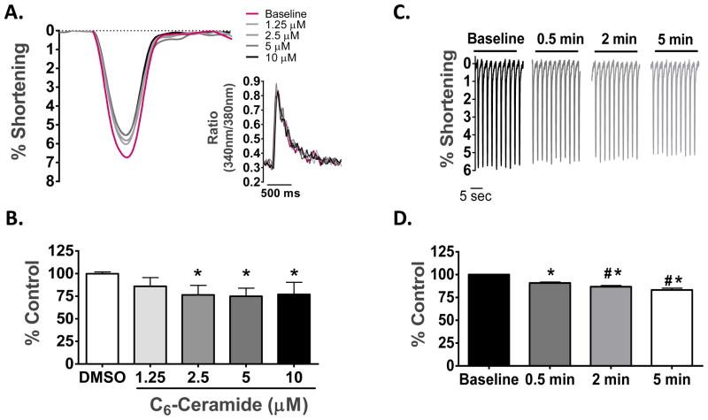
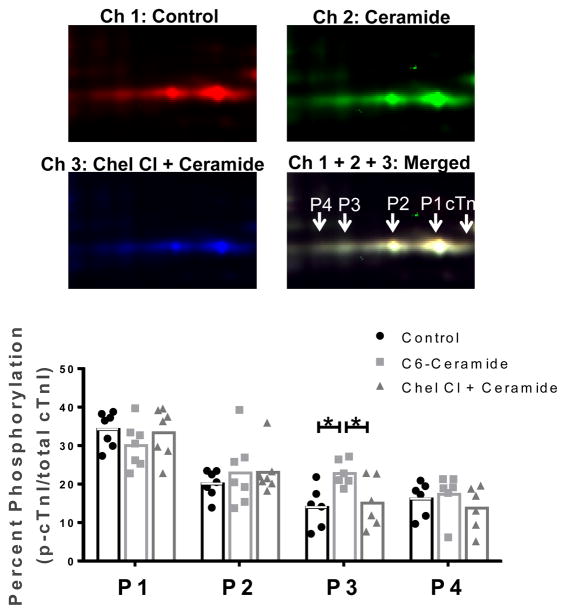
Although ceramide accumulation in the heart is considered a major factor in promoting apoptosis and cardiac disorders, including heart failure, lipotoxicity and ischemia-reperfusion injury, little is known about ceramide's role in mediating changes in contractility. In the present study, we measured the functional consequences of acute exposure of isolated field-stimulated adult rat cardiomyocytes to C6-ceramide. Exogenous ceramide treatment depressed the peak amplitude and the maximal velocity of shortening without altering intracellular calcium levels or kinetics. The inactive ceramide analog C6-dihydroceramide had no effect on myocyte shortening or [Ca(2+)]i transients. Experiments testing a potential role for C6-ceramide-mediated effects on activation of protein kinase C (PKC) demonstrated evidence for signaling through the calcium-independent isoform, PKCε. We employed 2-dimensional electrophoresis and anti-phospho-peptide antibodies to test whether treatment of the cardiomyocytes with C6-ceramide altered myocyte shortening via PKC-dependent phosphorylation of myofilament proteins. Compared to controls, myocytes treated with ceramide exhibited increased phosphorylation of myosin binding protein-C (cMyBP-C), specifically at Ser273 and Ser302, and troponin I (cTnI) at sites apart from Ser23/24, which could be attenuated with PKC inhibition. We conclude that the altered myofilament response to calcium resulting from multiple sites of PKC-dependent phosphorylation contributes to contractile dysfunction that is associated with cardiac diseases in which elevations in ceramides are present.


Conflict of interest statement

CONFLICT OF INTEREST

None

Figures

Fig. 1
Fig. 1. Western blot verifying efficient subcellular fractionation
Representative Western blot of GAPDH and Na+/K+ ATPase performed on samples taken during each step of the subcellular fractionation protocol (see Materials and Methods). The presence of GAPDH predominately within the cytosolic fraction and Na+/K+ ATPase only within the corresponding membrane fraction confirms proper subcellular fractionation.
Fig. 2
Fig. 2. Concentration and time-dependent effects of exogenous C6-ceramide treatment on contractile mechanics and [Ca2+]i in ventricular cardiomyocytes
(A) Overlay of unloaded cell shortening measurements from a single cardiomyocyte under field stimulation at 0.5 Hz at baseline and during treatment with sequentially increasing concentrations of C6-ceramide. Insert, overlay of simultaneous Ca2+-transient measures corresponding to each cell shortening measure. (B) The quantified change in the percentage of cell shortening relative to control following treatment with vehicle (DMSO) and each concentration of C6-ceramide. * p < 0.01 vs DMSO; n = 6. (C) Representative cell shortening measurements taken under steady-state pacing at baseline conditions and following 30 sec, 2 min and 5 min of exogenous 5 μM C6-ceramide treatment. ((D) The quantified change in the percentage of cell shortening relative to control during the time course for 5 μM C6-ceramide treatment. *p>0.05 vs. Baseline, #p<0.01 vs. 0.5 min, n = 8.
Fig. 3
Fig. 3. Effects of acute C6-ceramide treatment on contractile mechanics and [Ca2+]i in ventricular cardiomyocytes
(A) Overlay of representative unloaded cell shortening measurements with simultaneous Ca2+ transient measurements in a single cardiomyocyte under field stimulation (0.5 Hz) before (black) and after (red) exogenous treatment with 5 μM C6-ceramide. (B) Quantification of the contractile parameters (percent of shortening and maximal rates of contraction and relaxation) and (C), Ca2+ transient parameters (peak amplitude of the Fura-2 ratio and the time constant, τ, for the declining phase of the transient). *p<0.05 vs Control, ** p<0.001 vs Control, n=8.
Fig. 4
Fig. 4. Assessment of functional specificity using the non-bioactive sphingolipid C6-Dihydroceramide
(A) Quantification of contractile parameters (percent of shortening and maximal rates of contraction and relaxation) before treatment, after vehicle treatment (0.05% DMSO) and following treatment with 5 μM C6-dihydroceramide. (B) Quantification of Ca2+ transient parameters (peak amplitude of the Fura ratio (340 nm/380 nm) and the time constant, τ, for the declining phase of the transient) before treatment, after vehicle treatment and following treatment with 5 μM C6-dihydroceramide. n=5.
Fig. 5
Fig. 5. Attenuation of the functional effects of acute C6-ceramide treatment by inhibition of PKCs
(A) Quantification of contractile parameters (percent of shortening and maximal rates of contraction and relaxation) following treatment with 1 μM chelerythrine chloride and 1 μM chelerythrine chloride + 5 μM C6-ceramide. n=6 (B) Quantification of contractile parameters (percent of shortening and maximal rates of contraction and relaxation) following treatment with 5 μM Go6976 and 5 μM Go6976 + 5 μM C6-ceramide. *p<0.01 vs Control, n=6.
Fig. 6
Fig. 6. Subcellular fractionation of ventricular cardiomyocytes treated with C6-ceramide to assess translocation of the calcium-independent PKC isoforms
(A) Representative Western blots of the calcium-independent PKC isoforms in the cytosolic and membrane subcellular fractions of isolated cardiomyocytes in control cells, cells treated with 100 nM PMA, and cells treated with 10 μM C6-ceramide. (B) Quantification of the cytosolic and membrane subcellular fractions from control and C6-ceramide treated samples. White bars represent the amount of PKC in the cytosolic fraction and grey bars represent the amount of PKC within the membrane fraction. Each band density was normalized to the total protein within that lane. *p<0.05 vs Control, n=3.
Fig. 7
Fig. 7. The role of PKC in C6-ceramide-mediated effects on cTnI phosphorylation
cTnI, cardiac troponin I; P, phosphorylated cardiac troponin I. Top, images of the cTnI region of interest from Cy5 labeled control samples (channel 1), Cy2 labeled samples treated with 10 μM C6-ceramide (channel 2) and Cy3 labeled samples treated with 10 μM C6-ceramide in the presence of 1 μM Chelerytherine chloride (Channel 3) and the merged image (channel 1 + 2 + 3). Samples were pooled together and focused in the first dimension on a 18 cm nonlinear IPG 7–11 pH strip followed by standard 12% SDS-PAGE for the second dimension. Bottom, quantitative results for cTnI phosphorylation spots 1–4 in which each spot density is shown relative to the total density for all cTnI spots. *p<0.05 as indicated; n=7.
Fig. 8
Fig. 8. 2D-DIGE of myofilament protein phosphorylation for Tm, RLC, cTnT and cMyBP-C
Tm, tropomyosin; RLC, regulatory light chain; cTnT, cardiac troponin T, MyBP-C, myosin binding protein-C; P, phosphorylation, M, modification. A, Top, representative gel images for the region of interest including Tm, RLC and cTnT are shown in a merged image of control samples labeled with Cy3 (pseudo-colored green) and C6-ceramide treated samples labeled with Cy5 (pseudo-colored red). Bottom, Results from the quantification of phosphorylation sites from Tm, RLC and cTnT expressed as a percentage of all spot density for the protein of interest. B, Top, representative gel image for cMyBP-C shown as a merged image of control samples labeled with Cy3 and C6-ceramide treated samples labeled with Cy5. Bottom, Results from the quantification of modification sites on cMyBP-C expressed as a percentage of all spot density. *p<0.05 vs Control; n = 7.
Fig. 9
Fig. 9. Site-specific immunoblotting for cMyBP-C, Tm and cTnI phosphorylation
(A) Left, Representative images from Western blots using cMyBP-C phospho-specific antibodies against Ser-273, Ser-282 and Ser-302 in untreated samples and in samples treated with increasing doses of C6-ceramide. Right, Quantification of cMyBP-C phosphorylation levels in isolated cells treated with and without 10 μM C6-ceramide at Ser-273, Ser-282 and Ser302. GAPDH was used as a loading control. n=5 (B) Western blot quantification of isolated cells treated with and without 10 μM C6-ceramide using a phospho-Tm (Ser283) antibody. n=5 (C) Western blot quantification of isolated cells treated with and without 10 μM C6-ceramide using a phospho-cTnI (Ser23/24) antibody. n=6; *p<0.05 vs Control.
Fig. 10
Fig. 10. Schematic diagram of the mechanism by which ceramide leads to negative inotropy
Accumulation of ceramide within the cardiomyocyte results in activation of PKCε, leading to phosphorylation of the myofilament proteins cTnI and cMyBP-C. These post-translational modifications within the sarcomere alter the basal function and cause a reduction in the ability of the contractile mechinary to generate force.

Similar articles

Cited by

References

    1. Ayaz-Guner S, Zhang J, Li L, Walker JW, Ge Y. In vivo phosphorylation site mapping in mouse cardiac troponin I by high resolution top-down electron capture dissociation mass spectrometry: Ser22/23 are the only sites basally phosphorylated. Biochemistry. 2009;48:8161–8170. doi: 10.1021/bi900739f. - DOI - PMC - PubMed
    1. Bielawska A, Crane HM, Liotta D, Obeid LM, Hannun YA. Selectivity of ceramide-mediated biology. Lack of activity of erythro-dihydroceramide. J Biol Chem. 1993;268:26226–26232. - PubMed
    1. Bowman JC, Steinberg SF, Jiang T, Geenen DL, Fishman GI, Buttrick PM. Expression of protein kinase C beta in the heart causes hypertrophy in adult mice and sudden death in neonates. J Clin Invest. 1997;100:2189–2195. doi: 10.1172/JCI119755. - DOI - PMC - PubMed
    1. Brenner B. Effect of Ca2+ on cross-bridge turnover kinetics in skinned single rabbit psoas fibers: implications for regulation of muscle contraction. Proc Natl Acad Sci U S A. 1988;85:3265–3269. - PMC - PubMed
    1. Brutsaert DL, Sys SU. Relaxation and diastole of the heart. Physiol Rev. 1989;69:1228–1315. - PubMed

Publication types

LinkOut - more resources