. 2014 Dec 5:281:124-34.
doi: 10.1016/j.neuroscience.2014.09.055. Epub 2014 Oct 2.

Effect of status epilepticus and antiepileptic drugs on CYP2E1 brain expression

Affiliations

Effect of status epilepticus and antiepileptic drugs on CYP2E1 brain expression

B Boussadia et al. Neuroscience. .

Abstract

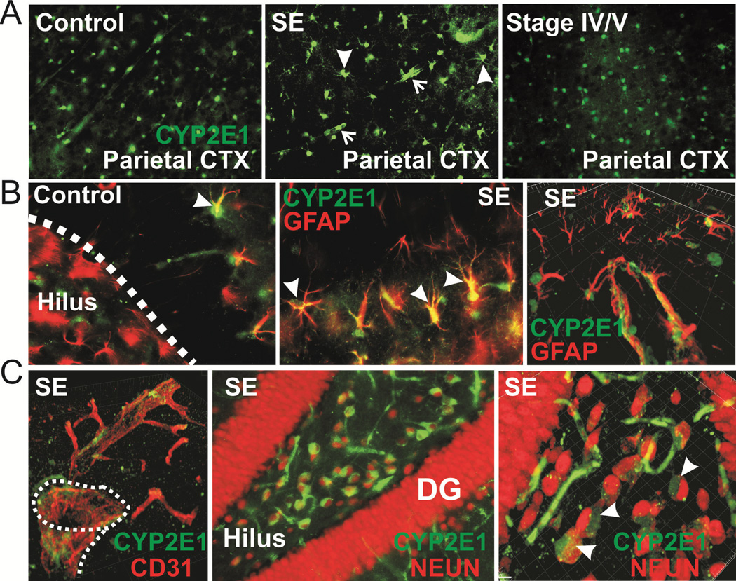
P450 metabolic enzymes are expressed in the human and rodent brain. Recent data support their involvement in the pathophysiology of epilepsy. However, the determinants of metabolic enzyme expression in the epileptic brain are unclear. We tested the hypothesis that status epilepticus (SE) or exposure to phenytoin or phenobarbital affects brain expression of the metabolic enzyme CYP2E1. SE was induced in C57BL/6J mice by systemic kainic acid. Brain CYP2E1 expression was evaluated 18-24h after severe SE by immunohistochemistry. Co-localization with neuronal nuclei (NEUN), glial fibrillary acidic protein (GFAP) and CD31 was determined by confocal microscopy. The effect of phenytoin, carbamazepine and phenobarbital on CYP2E1 expression was evaluated in vivo or by using organotypic hippocampal cultures in vitro. CYP2E1 expression was investigated in brain resections from a cohort of drug-resistant epileptic brain resections and human endothelial cultures (EPI-EC). Immunohistochemistry showed an increase of CYP2E1 expression limited to hippocampal CA2/3 and hilar neurons after severe SE in mice. CYP2E1 expression was also observed at the astrocyte-vascular interface. Analysis of human brain specimens revealed CYP2E1 expression in neurons and vascular endothelial cells (EC). CYP2E1 was expressed in cultured human EC and over-expressed by EPI-EC. When analyzing the effect of drug exposure on CYP2E1 expression we found that, in vivo or in vitro, ethanol increased CYP2E1 levels in the brain and liver. Treatment with phenytoin induced localized CYP2E1 expression in the brain whereas no significant effects were exerted by carbamazepine or phenobarbital. Our data indicate that the effect of acute SE on brain CYP2E1 expression is localized and cell specific. Exposure to selected anti-epileptic drugs could play a role in determining CYP2E1 brain expression. Additional investigation is required to fully reproduce the culprits of P450 enzyme expression as observed in the human epileptic brain.

Keywords: CYP2E1; biotransformation; drug exposure; status epilepticus.


Conflict of interest statement

Conflict of Interest

All authors have no conflict of interest to declare. The authors have no relationships with organizations that could inappropriately influence, or be perceived to influence, the presented work.

Figures

Figure 1
Figure 1. Increased CYP2E1 expression is localized in the hippocampus after severe SE
A) Control mice display CYP2E1 staining in glial cells and sporadic vessels throughout the hippocampus (see also co-localization in Figure 2). CYP2E1 immunoreactivity was negligible in the CA1. A number of hilar and CA2/3 neurons were positive for CYP2E1 (see quantification in C). Mice experiencing severe SE display increased immunoreactivity specifically in the CA2/3 regions and in the hilar portion of the dentate gyrus (neurons and reactive astrocytes indicated by arrowheads and vascular staining indicated by arrows). B–B1) Example of RGB stacks images utilized for the quantification of CYP2E1 fluorescence. CYP2E1 is expressed as % pixels to control mean (see Methods). C–C1) Note the significant increase in the CA2/3 regions of SE animals, while in the hilar/DG portion a trend increase was measured. Expression of CYP2E1 in animals experiencing Stage IV/V Racine changes did not increase. Data are indicated by a box plot as mean ± SE (ANOVA).
Figure 2
Figure 2. CYP2E1 colocalization with GFAP, NEUN and CD31
A) The pattern of CYP2E1 cortical expression was slightly modified after severe SE as compared to control. A number of CYP2E1+ vessels were visible after severe SE only (arrows). In mice experiencing behavioral stage IV–V CYP2E1 immunoreactivity was identical to control. B) CYP2E1 co-localized with activated astrocytes (GFAP+; arrowheads). An example of hippocampal staining is provided (control and SE). C) CYP2E1 expression was associated with CD31+ vessels in the cortex and hippocampus after SE. Examples of confocal 3D reconstructions (CD31 and NEUN) are provided.
Figure 3
Figure 3. Evaluation of CYP2E1 expression in the human TLE brain tissues and endothelial cells
A) CYP2E1 staining was observed at the vasculature, astrocytes (GFAP+) and neurons (NEUN+). Images depict examples obtained from gliotic regions. B) Example of CYP2E1 expression in a brain specimens obtained from a patient receiving phenytoin (see also Table 1). C–C1) CYP2E1 is expressed in brain endothelial cells (HBMEC and EPI-EC; see Methods for details) and it is over-expressed in EPI-EC (n = 4, see Table 1). While CYP2E1 levels were increased in EPI-EC as compared to HBMEC, variable CYP2E1 levels were found within the pool of EPI-EC analyzed. Data are indicated by a box plot as mean ± SE (ANOVA).
Figure 4
Figure 4. Immunohistochemistry of CYP2E1 brain expression after in vivo drug exposure
A–A1) Treatment with ethanol, a known CYP2E1 inducer, increased enzyme brain immunostaining. Note the appearance of distinct vascular signal in the cortical and hippocampal areas (arrowheads). Interestingly, in vivo treatment with phenytoin also increased CYP2E1 staining. Treatment with phenobarbital provoked minor effects on CYP expression, mostly confined to the hippocampus. B) Images depict examples of cortical RGB stacks used for CYP2E1 quantification (see also Methods). Note the increased on CYP2E1 immunoreactivity in animals after treatment with phenytoin. Data are indicated by a box plot as mean ± SE (ANOVA).
Figure 5
Figure 5. Western blot analysis of CYP2E1 brain expression after in vivo drug exposure
A) CYP2E1 tissue levels were increased (significant or trend) in the cortex after ethanol or phenytoin treatments. B) In the hippocampus phenytoin determined an increase of CYP2E1 expression. C) Ethanol, and not phenytoin or phenobarbital, determined a trend increase in CYP2E1 expression in the liver. D) Examples of western blots are shown. Data are indicated by a box plot (mean ± SE; ANOVA). EE3) In vivo, carbamazepine treatment did not affect brain or hepatic CYP2E1 levels. Note that treatment with vehicle (PEG 400) alone was associated with increased enzyme expression.
Figure 6
Figure 6. Western blot analysis of CYP2E1 brain expression after in vitro drug exposure
A) Data are relative to n = 5 in vitro OHC (1 ETOH; 2 Phenytoin; 2 Phenobarbital; see Methods). Phenytoin and ethanol induced CYP2E1 expression in organotypic hippocampal cultures. Box plot depicts the CYP2E1 differences between treatments while x–y plots in B, C and D depict CYP2E1 levels in function of drug dosage. Each value refers to CYP2E1 quantification from 16 slices pulled together from 4 wells (n = 8 pups). Data are normalized by respective OHC control (100%; see Methods for details). Linear regression was used to analyze data in B, C and D (Pearson’s coefficient).

References

    1. Abbott NJ, Khan EU, Rollinson CM, Reichel A, Janigro D, Dombrowski SM, Dobbie MS, Begley DJ. Drug resistance in epilepsy: the role of the blood-brain barrier. NovartisFoundSymp. 2002;243:38–47. - PubMed
    1. Bankstahl JP, Hoffmann K, Bethmann K, Loscher W. Glutamate is critically involved in seizure-induced overexpression of P-glycoprotein in the brain. Neuropharmacology. 2008;54:1006–1016. - PubMed
    1. Bankstahl M, Bankstahl JP, Loscher W. Pilocarpine-induced epilepsy in mice alters seizure thresholds and the efficacy of antiepileptic drugs in the 6-Hertz psychomotor seizure model. Epilepsy research. 2013;107:205–216. - PubMed
    1. Bauer B, Hartz AM, Fricker G, Miller DS. Pregnane X receptor up-regulation of P-glycoprotein expression and transport function at the blood-brain barrier. MolPharmacol. 2004;66:413–419. - PubMed
    1. Bauer B, Yang X, Hartz AM, Olson ER, Zhao R, Kalvass JC, Pollack GM, Miller DS. In vivo activation of human pregnane X receptor tightens the blood-brain barrier to methadone through P-glycoprotein up-regulation. MolPharmacol. 2006;70:1212–1219. - PubMed

MeSH terms

LinkOut - more resources