Case Reports
. 2014 Dec;16(4):821-44.
doi: 10.1007/s12017-014-8331-9. Epub 2014 Oct 4.

A diagnostic approach for cerebral palsy in the genomic era

Affiliations
Case Reports

A diagnostic approach for cerebral palsy in the genomic era

Ryan W Lee et al. Neuromolecular Med. 2014 Dec.

Abstract

An ongoing challenge in children presenting with motor delay/impairment early in life is to identify neurogenetic disorders with a clinical phenotype, which can be misdiagnosed as cerebral palsy (CP). To help distinguish patients in these two groups, conventional magnetic resonance imaging of the brain has been of great benefit in "unmasking" many of these genetic etiologies and has provided important clues to differential diagnosis in others. Recent advances in molecular genetics such as chromosomal microarray and next-generation sequencing have further revolutionized the understanding of etiology by more precisely classifying these disorders with a molecular cause. In this paper, we present a review of neurogenetic disorders masquerading as cerebral palsy evaluated at one institution. We have included representative case examples children presenting with dyskinetic, spastic, and ataxic phenotypes, with the intent to highlight the time-honored approach of using clinical tools of history and examination to focus the subsequent etiologic search with advanced neuroimaging modalities and molecular genetic tools. A precise diagnosis of these masqueraders and their differentiation from CP is important in terms of therapy, prognosis, and family counseling. In summary, this review serves as a continued call to remain vigilant for current and other to-be-discovered neurogenetic masqueraders of cerebral palsy, thereby optimizing care for patients and their families.


Conflict of interest statement

CONFLICTS OF INTEREST

The corresponding author is a paid member of the drug monitoring committee for BlueBirdBio, Inc. There are no other conflicts of interest.

Figures

Fig. 1
Fig. 1
Diagnostic Evaluation of the Child with Cerebral Palsy.
Fig. 2
Fig. 2
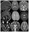
Masqueraders of dyskinetic CP. a, Axial T2-weighted image of a 5-year-old child with panthothenate kinase associated neurodegeneration and severe, generalized dystonia shows a bilateral, symmetric hyperintense signal abnormalities within the globus pallidi surrounded by rings of low signal intensity; b, Midsagittal T1- and c, Coronal T2-weighted images of a 12-month-old infant with pontocerebellar hypoplasia type 2 due to TSEN54 mutation, progressive microcephaly and dyskinetic movement disorders demonstrates a small cerebellum with enlarged intrafoliar spaces (the cerebellar hemispheres are more affected compared to the cerebellar vermis), a reduction in size of the pons, a delayed myelination and a cerebral atrophy; d, Axial T2-weighted image of a 10-years-old child with Wilson disease, severe dystonia and a bilateral Kayser-Fleischer ring reveals a bilateral, symmetric hyperintense signal within the putamina and caudate nuclei; e, Axial T2-weighted image of a 6-year-old boy with aromatic acid decarboxylase deficiency, dystonia, oculogyric crisis and global developmental delay shows a normal brain anatomy; f, Axial T2-weighted image of a 7-month-old girl with glutaric aciduria type 1, macrocephaly and severe dystonia demonstrates a bilateral, symmetric hyperintense signal in and atrophy of the putamina and caudate nuclei; g, Axial T2-weighted image of a 4-year-old girl with Segawa disease and progressive dystonia with diurnal fluctuations presents normal brain anatomy
Fig. 3
Fig. 3
Masqueraders of bilateral spastic CP. a, Axial T2-weighted image of a 1-day-old newborn with Miller-Dieker syndrome due to LIS1 mutation shows a lissencephalic brain with a smooth and abnormally thick cerebral cortex; the child developed severe epileptic seizures, bilateral spasticity and microcephaly and did not achieve almost any developmental milestones; b, Axial T2-weighted image of a 12-month-old infant with Alexander disease, progressive macrocephaly, regression and bilateral spasticity reveals symmetrical hyperintense signal abnormalities in the bilateral frontal white matter and arcuate fibers, caudate nuclei and putamina; c, Axial T2-weighted image of a 3-year-old girl with a “double cortex”, severe epileptic seizures and mild, bilateral spasticity demonstrates bilateral, subcortical heterotopic bands and ventriculomegaly; d, Axial T2-weighted image of a 3-month-old male infant with Pelizaeus-Merzbacher disease, bilateral nystagmus and progressive, bilateral spasticity shows complete absence of myelinated white matter (all the white matter appears T2-hyperintense); e, Axial T2-weighted image of a 6-month-old infant with Krabbe disease, irritability, progressive bilateral spasticity and regression reveals hyperintense signal abnormalities within the lateral/dorsal extension of the middle cerebellar peduncles (white arrows), dentate nuclei (white arrow heads) and periventricular white matter with predominant involvement of the posterior regions (not shown); f, Axial T2-weighted image of a 4-year-old child with bilateral open-lip schizencephaly, seizures and bilateral spasticity demonstrates bilateral clefts communicating with the lateral ventricles and outlined by dysplastic gray matter
Fig. 4
Fig. 4
Masqueraders of unilateral spastic CP. a, Axial T2-weighted image of a 4-year-old child with isolated left hemimegalencephaly, seizures and right spastic hemiparesis shows enlargement of the left cerebral hemisphere with dilatation of the left lateral ventricle and hyperintense signal abnormality of the left periventricular white matter; b, Axial T2-weighted image of a 3-year-old child with right sided open-lip schizencephaly, focal epileptic seizures and left sided spastic hemiparesis reveals a cleft within the right parietal lobe communicating with the lateral ventricle and outlined by dysplastic gray matter
Fig. 5
Fig. 5
Masqueraders of ataxic CP. a, Midsagittal T1-weighted image of a 2-year-old child with Dandy-Walker malformation and cerebellar ataxia shows hypoplasia of the cerebellar vermis, which is elevated and upward rotated (white arrows), and cystic dilatation of the fourth ventricle; b, Axial and c, Coronal T2-weighted images of a 3 year-old-girl with 4H syndrome (Hypomyelination, Hypodontia and Hypogonadotropic Hypogonadism) and ataxia reveals only few areas of low signal in the white matter (posterior limb of the internal capsule and optic radiation) compatible with hypomyelination and cerebellar atrophy; d, Midsagittal T1- and e, Axial T2-weihgted images of a 3-year-old child with Salla disease and ataxia demonstrates severe vermian atrophy, a thin corpus callosum and diffuse hyperintensity of the white matter as a sign of hypomyelination; f, Axial T2-weighted image at the level of the pontomesencephalic junction of a 2-year-old child with Joubert syndrome, ataxia, ocular motor apraxia and global developmental delay shows the pathognomonic ‘molar tooth sign’ characterized by a deepened interpeduncular fossa, elongated, thickened and horizontally orientated superior cerebellar peduncles (white arrows) and vermian hypo-dysplasia; g, Axial T2-weighted image of a 8-year-old child with rhombencephalosynapsis and ataxia reveals fused cerebellar hemispheres without intervening vermis, abnormal, transverse orientation of cerebellar folia and mild dilatation of the temporal horns of the lateral ventricles; h, Coronal and i, Axial T2-weighted images of a 3.5-year-old child with late-infantile neuronal ceroid lipofuscinoses, ataxia and myoclonic seizures demonstrates moderate global cerebellar atrophy, mild cerebral atrophy and symmetric hyperintensity of the periventricular white matter (images d, e, h and i are reprinted with permission from Poretti A et al., Differential diagnosis of cerebellar atrophy in childhood, Eur J Paediatr Neurol, 2008)

References

    1. Accardo PJCAJ. Capute & Accardo’s neurodevelopmental disabilities in infancy and childhood. Baltimore: Paul H. Brookes Pub; 2008.
    1. Ancel PY, Livinec F, Larroque B, Marret S, Arnaud C, Pierrat V, et al. Cerebral palsy among very preterm children in relation to gestational age and neonatal ultrasound abnormalities: the EPIPAGE cohort study. Pediatrics. 2006;117(3):828–835. doi: 10.1542/peds.2005-0091. - DOI - PubMed
    1. Andersen GL, Irgens LM, Haagaas I, Skranes JS, Meberg AE, Vik T. Cerebral palsy in Norway: prevalence, subtypes and severity. Eur J Paediatr Neurol. 2008;12(1):4–13. doi: 10.1016/j.ejpn.2007.05.001. - DOI - PubMed
    1. Apetauerova D, Schirmer CM, Shils JL, Zani J, Arle JE. Successful bilateral deep brain stimulation of the globus pallidus internus for persistent status dystonicus and generalized chorea. J Neurosurg. 2010;113(3):634–638. doi: 10.3171/2010.1.JNS091127. - DOI - PubMed
    1. Ashwal S, Russman BS, Blasco PA, Miller G, Sandler A, Shevell M, et al. Practice parameter: diagnostic assessment of the child with cerebral palsy: report of the Quality Standards Subcommittee of the American Academy of Neurology and the Practice Committee of the Child Neurology Society. Neurology. 2004;62(6):851–863. - PubMed

Publication types

MeSH terms

Substances