. 2014 Nov 15;193(10):5229-39.
doi: 10.4049/jimmunol.1400753. Epub 2014 Oct 3.

Fibrocytes are not an essential source of type I collagen during lung fibrosis

Affiliations

Fibrocytes are not an essential source of type I collagen during lung fibrosis

Kathryn R Kleaveland et al. J Immunol. .

Abstract

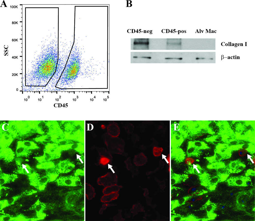
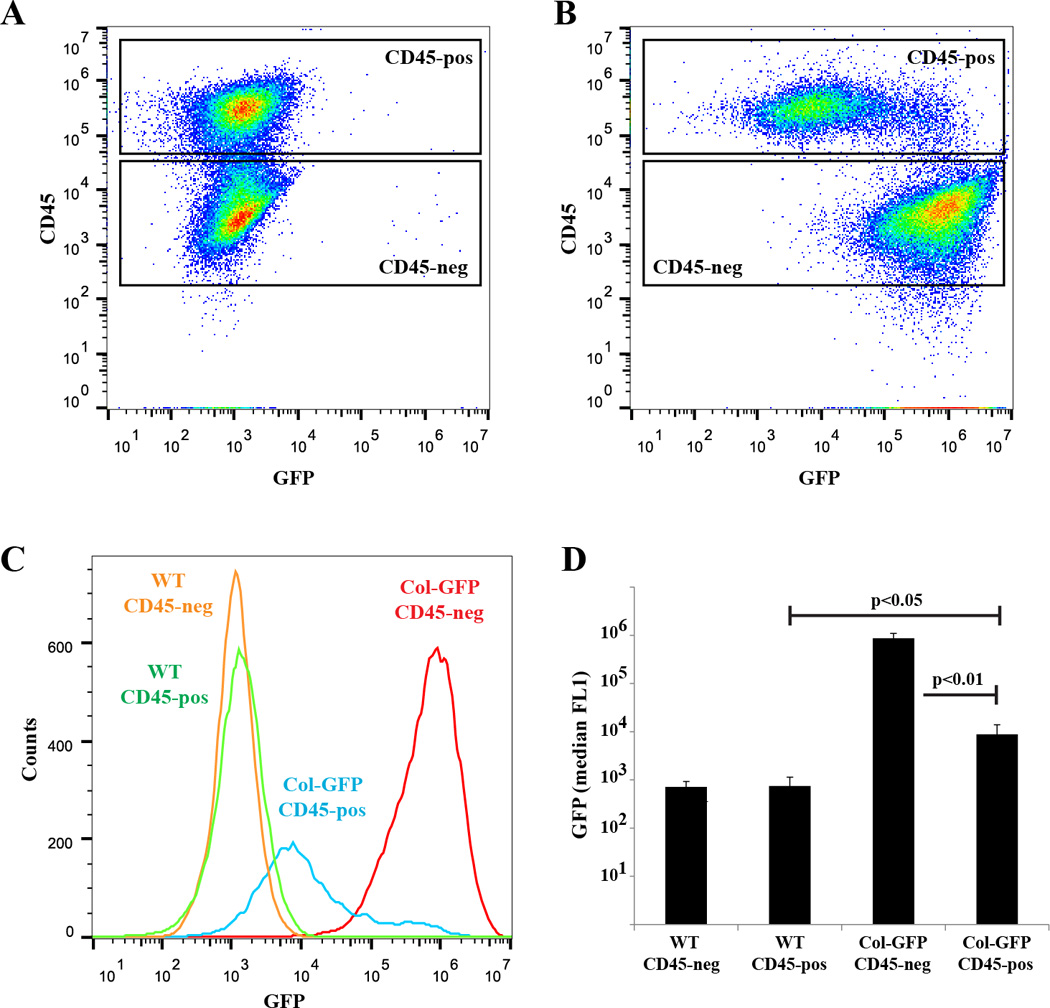
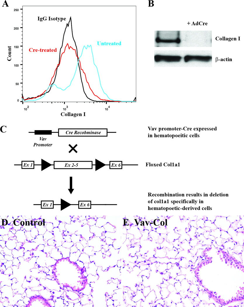
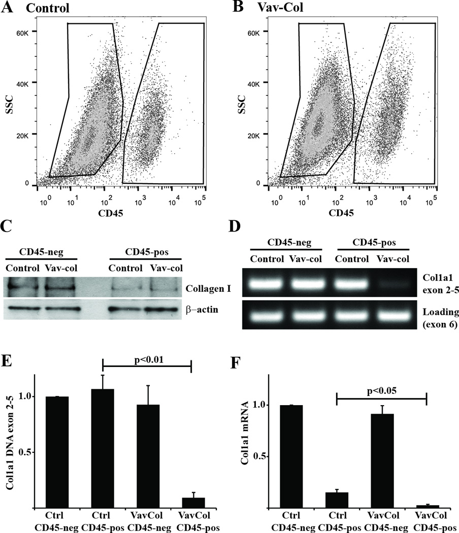
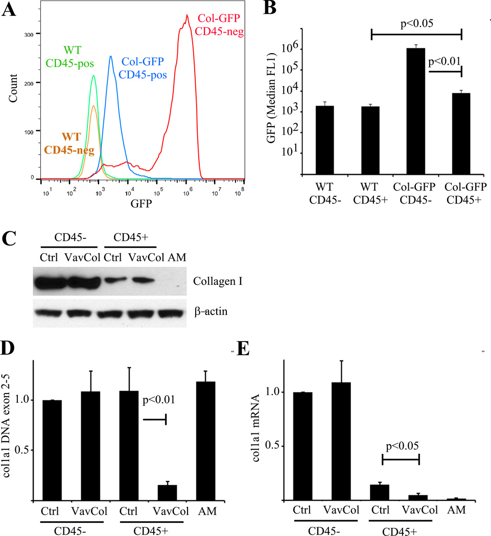
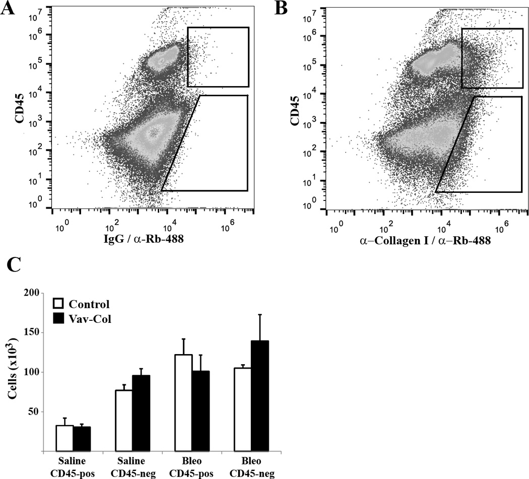
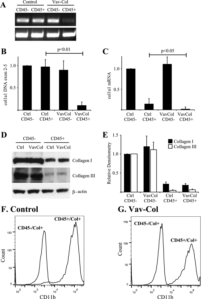
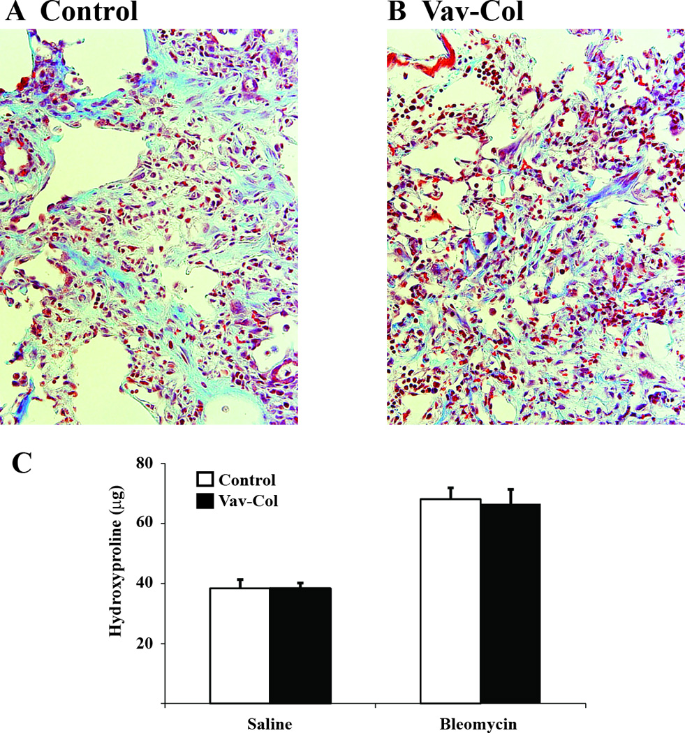
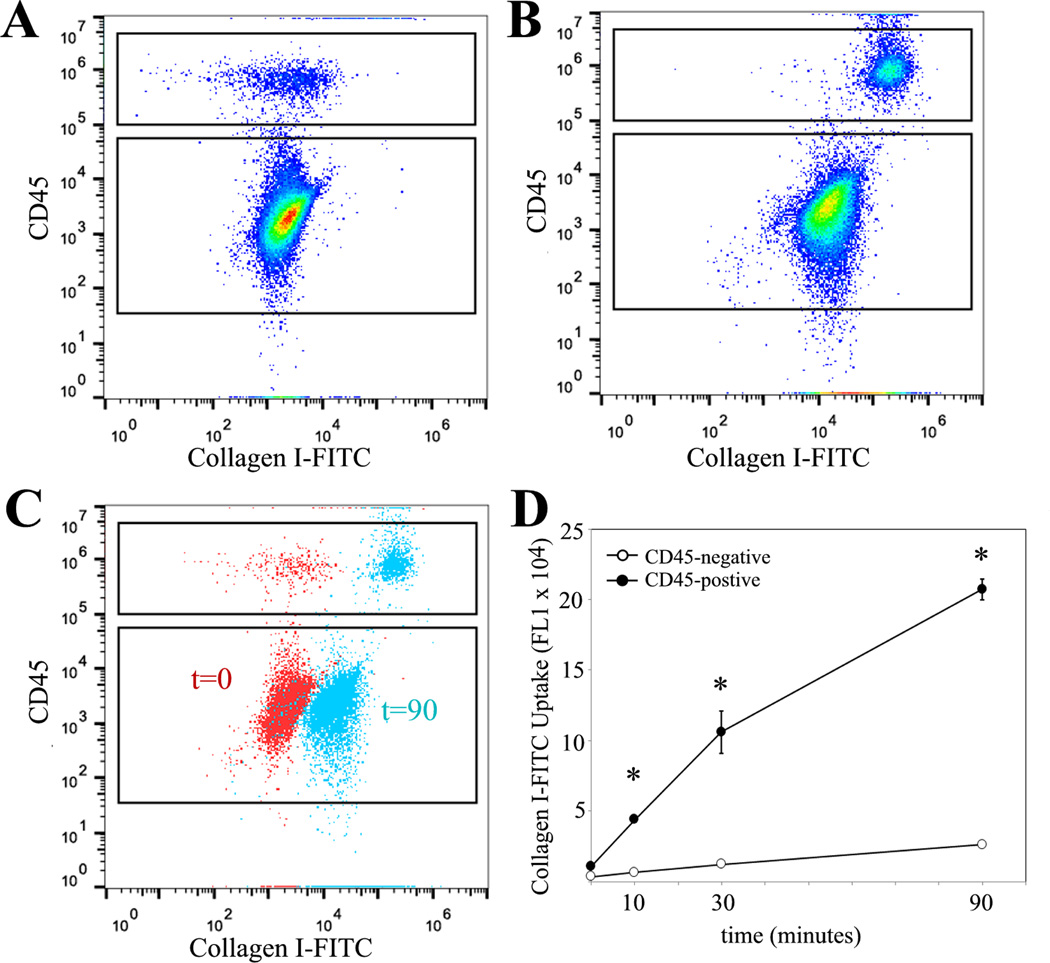
Progressive fibrosis involves accumulation of activated collagen-producing mesenchymal cells. Fibrocytes are hematopoietic-derived cells with mesenchymal features that potentially have a unique and critical function during fibrosis. Fibrocytes have been proposed as an important direct contributor of type I collagen deposition during fibrosis based largely on fate-mapping studies. To determine the functional contribution of hematopoietic cell-derived type I collagen to fibrogenesis, we use a double-transgenic system to specifically delete the type I collagen gene across a broad population of hematopoietic cells. These mice develop a robust fibrotic response similar to littermate genotype control mice injured with bleomycin indicating that fibrocytes are not a necessary source of type I collagen. Using collagen-promoter GFP mice, we find that fibrocytes express type I collagen. However, fibrocytes with confirmed deletion of the type I collagen gene have readily detectable intracellular type I collagen indicating that uptake of collagen from neighboring cells account for much of the fibrocyte collagen. Collectively, these results clarify several seemingly conflicting reports regarding the direct contribution of fibrocytes to collagen deposition.


Conflict of interest statement

The authors have no conflicting financial interests.

Figures

Figure 1
Figure 1. In vitro-derived lung mesenchymal cells contain a CD45-positive / collagen I-positive population
A) Primary lung mesenchymal cells from wild-type mice were stained for CD45 and analyzed by flow cytometry. The majority of cells are CD45-negative but a substantial subset are CD45-positive. B) The CD45-negative and positive populations were FACS sorted and analyzed by immunoblot and compared to alveolar macrophages as a negative control. Both CD45-positive and negative cells contained type I collagen protein but CD45-negative cells contained significantly more. Wild-type cells were co-stained for type I collagen (C), CD45 (D), and merged (E) demonstrating weak type I collagen staining within CD45-positive cells (200×).
Figure 2
Figure 2. In vitro-derived lung fibrocytes express type I collagen
A&B) Primary lung mesenchymal cells from wild-type (A) and Col-GFP (B) mice were isolated and analyzed for expression of GFP and CD45 by flow cytometry. C) Histogram overlay of GFP expression by CD45-positive and negative lung mesenchymal cells. D) Quantification of GFP expression demonstrating greater GFP expression in CD45-negative fibroblasts (p<0.01), but significant GFP within CD45-positive fibrocytes from Col-GFP mice compared to wild-type cells (p<0.05), n=3.
Figure 3
Figure 3. Generation and of Mice with Deletion of Col1a1 within Hematopoietic Cells
A&B) Primary lung mesenchymal cells from floxed col1a1 (col1a1fl/fl) mice were treated with adenovirus expressing Cre recombinase in vitro leading to loss of type I collagen by flow cytometry (A) and immmunoblot (B). C) Hematopoietic cell-specific and permanent deletion of col1a1 is achieved using double transgenic mice carrying the Vav-Cre transgene crossed to col1a1fl/fl mice (Vav-Col). The Vav promoter yields Cre expression specifically within hematopoietic cells resulting in permanent deletion col1a1 within hematopoietic cells and their derivatives. D&E) Uninjured lungs from Vav-Col mice (E) have normal histology compared to littermate genotype control mice lacking either the Vav-Cre or floxed col1a1 allele (D), H&E (200×).
Figure 4
Figure 4. Fibrocytes from Vav-Col mice contain type I collagen protein even in the absence of type I collagen gene
A&B) Primary lung mesenchymal cells from uninjured control (A) and Vav-Col (B) mice were FACS sorted for CD45-positive (pos) and negative (neg) populations. The Vav-Col and littermate control mice had similar proportions of CD45-pos and -neg cells. C) Immunoblot for type I collagen. CD45-neg cells have more type I collagen than CD45-pos cells. CD45-pos cells from control and Vav-Col mice have similar levels of type I collagen protein. D) PCR of DNA isolated from control and Vav-Col CD45-pos and -neg cells. Primers within the floxed region of col1a1 demonstrate absence of exons 2–5 in Vav-Col CD45-pos cells and intact col1a1 in CD45-neg cells and cells from control mice. Primers outside of the floxed region (in exon 6) were used as a loading control. E&F) CD45-pos cells from Vav-Col mice have less col1a1 exons 2–5 DNA (p<0.01) by qPCR (E) and less col1a1 mRNA by qPCR (F) (p<0.05) compared to CD45-pos cells from control mice, n=4.
Figure 5
Figure 5. In vitro-derived fibrocytes from bleomycin injured mice express
A&B) Two weeks after intratracheal bleomycin injury, minced lung outgrowth mesenchymal cells from wild-type and Col-GFP mice were isolated and analyzed by flow cytometry. A) Histogram overlay of GFP expression by CD45-postive (CD45+) and CD45-negative (CD45−) cells. B) Quantification of GFP expression demonstrating greater GFP expression in CD45- fibroblasts (p<0.01), but significant GFP within CD45+ fibrocytes from Col-GFP mice compared to wild-type cells (p<0.05), n=4. C–E) Two weeks after bleomycin minced lung outgrowth mesenchymal cells from control and Vav-Col mice were sorted for CD45 and compared to alveolar macrophage (AM) control. C) Immunoblot for type I collagen demonstrating less type I collagen within CD45+ cells but similar amounts of type I collagen between CD45+ cells from control mice and Vav-Col mice. D) DNA qCR for floxed col1a1 exons 2–5 DNA demonstrates CD45+ cells from Vav-Col mice have robust recombination compared to other samples (p<0.01), n=4. E) Vav-Col mice have loss of col1a1 mRNA compared to CD45+ cells from control mice by qPCR (p<0.05), n=4.
Figure 6
Figure 6. Vav-Col mice have similar numbers of fibrocytes compared to genotype control mice after bleomycin
A&B) Single cell whole lung preparation from control and Vav-Col mice were analyzed by flow cytometry for CD45 and type I collagen (B) or isotype control for collagen staining (A). C) Bleomycin injury leads to increased numbers of collagen-positive cells. Bleomycin-injured Vav-Col mice have similar numbers of collagen-positive fibrocytes compared to littermate control mice (n=4).
Figure 7
Figure 7. Hematopoietic cell deletion of col1a1 does not affect fibrocyte type I collagen protein levels in vivo
A) DNA was isolated from FACS sorted CD45-positive (CD45+)/collagen I-positive and CD45-negative (CD45−)/collagen I-positive cells from Vav-Col and control mice two weeks after bleomycin injury. PCR demonstrates loss of col1a1 exons 2–5 in CD45+/collagen I-positive fibrocytes from Vav-Col mice compared to intact col1a1 in cells from genotype control mice and CD45− cells from Vav-Col mice. Primers outside the floxed region (exon 6) are used as a loading control. B) qPCR DNA analysis for col1a1 exons 2–5 demonstate significant recomination within CD45+ cells from Vav-Col mice, n=4. C) qPCR analysis for col1a1 mRNA. CD45+ cells have less col1a1 mRNA than CD45− cells. CD45+ cells from Vav-Col mice have near complete absense of col1a1 mRNA compared to CD45+ cells from genotype control mice (p<0.05, n=4). D&E) Immunoblot (D) demonstrates similar levels of type I and type III collagen within CD45+ cells from littermate control and Vav-Col mice quantified by densitometry (E), n=4. F&G) CD45+/collagen I-positive (CD45+/Col+) fibrocytes from littermate control (F) and Vav-Col (G) mice also co-express CD11b compared to CD45−/collagen I-positive (CD45−/Col+) fibroblasts.
Figure 8
Figure 8. Mice with hematopoietic cell deletion of col1a1 are not protected from bleomycin-induced lung fibrosis
A&B) Trichrome stained lung sections from littermate control (A) and Vav-Col (B) mice three weeks after bleomycin injury demonstrate robust fibrosis in both (400×). C) Hydroxyproline assay from lungs of control and Vav-Col mice three weeks after intratracheal saline or bleomycin demonstrate similar induction of fibrosis (n=4–10).
Figure 9
Figure 9. Lung fibrocytes readily take up FITC-conjugated collagen in vitro
A&B) Flow cytometry of primary mesenchymal cells for CD45 and FITC without addition of FITC-conjugated collagen I (t=0) (A) or 90 minutes after addition of FITC-conjugated (50 ug/mL) collagen I to the culture media (t=90) (B). C) Overlay of t=0 and t=90 plots demonstrating greater uptake of FITC-conjugated collagen I in CD45-positive cells. D) Timecourse of FITC-conjugated collagen I uptake demonstrating greater uptake by CD45-positive cells compared to CD45-negative cells (*p<0.01).

References

    1. American Thoracic Society. Idiopathic pulmonary fibrosis: diagnosis and treatment. International consensus statement. American Thoracic Society (ATS), and the European Respiratory Society (ERS) Am J Respir Crit Care Med. 2000;161:646–664. - PubMed
    1. American Thoracic Society/European Respiratory Society International Multidisciplinary Consensus Classification of the Idiopathic Interstitial Pneumonias. This joint statement of the American Thoracic Society (ATS), and the European Respiratory Society (ERS) was adopted by the ATS board of directors, June 2001 and by the ERS Executive Committee, June 2001. Am J Respir Crit Care Med. 2002;165:277–304. - PubMed
    1. Selman M, King TE, Pardo A. Idiopathic pulmonary fibrosis: prevailing and evolving hypotheses about its pathogenesis and implications for therapy. Ann Intern Med. 2001;134:136–151. - PubMed
    1. Thannickal VJ, Toews GB, White ES, Lynch JP, 3rd, Martinez FJ. Mechanisms of pulmonary fibrosis. Annu Rev Med. 2004;55:395–417. - PubMed
    1. Meltzer EB, Noble PW. Idiopathic pulmonary fibrosis. Orphanet J Rare Dis. 2008;3:8. - PMC - PubMed

Publication types

MeSH terms