. 2015 Jan:11:27-36.
doi: 10.1016/j.actbio.2014.09.032. Epub 2014 Oct 2.

Silk microfiber-reinforced silk hydrogel composites for functional cartilage tissue repair

Affiliations

Silk microfiber-reinforced silk hydrogel composites for functional cartilage tissue repair

Supansa Yodmuang et al. Acta Biomater. 2015 Jan.

Abstract

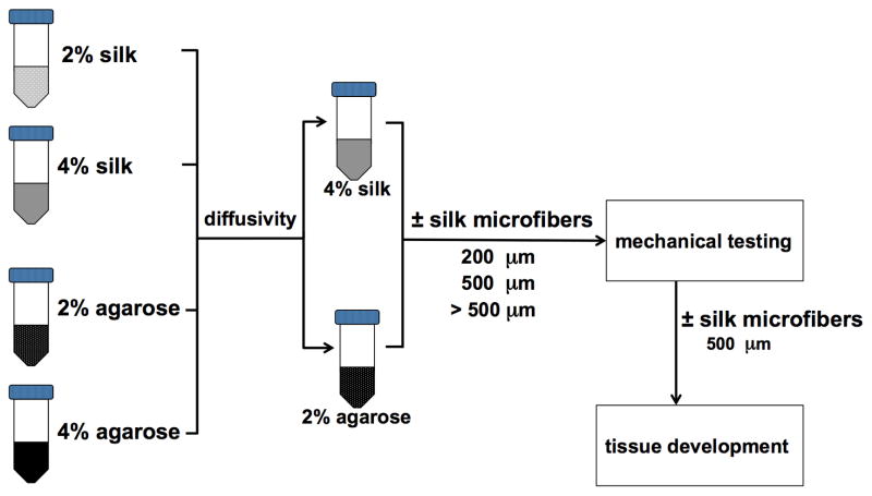
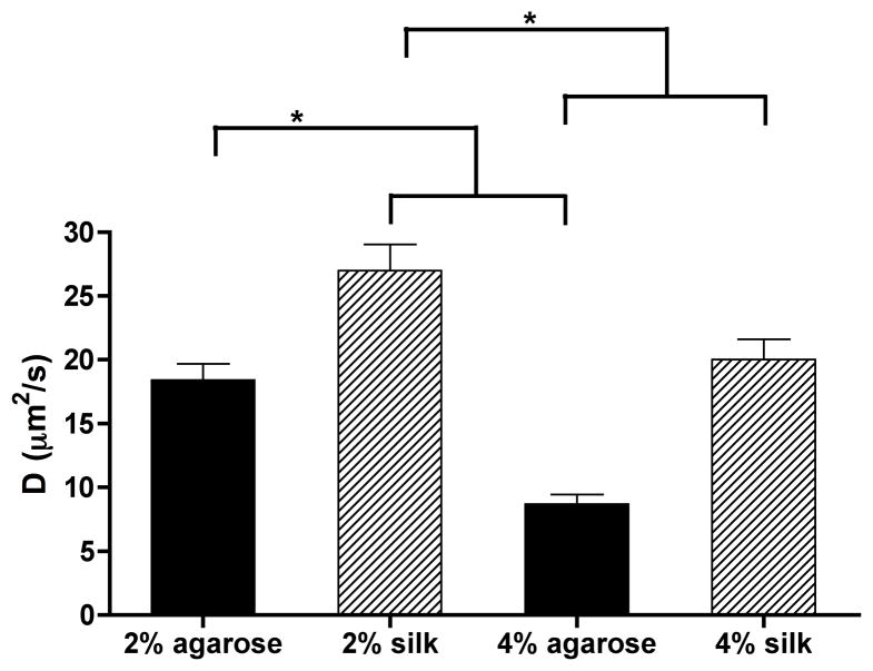
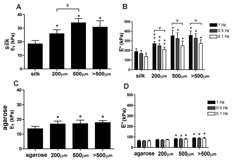
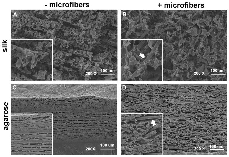
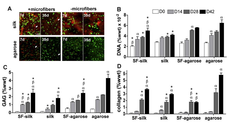
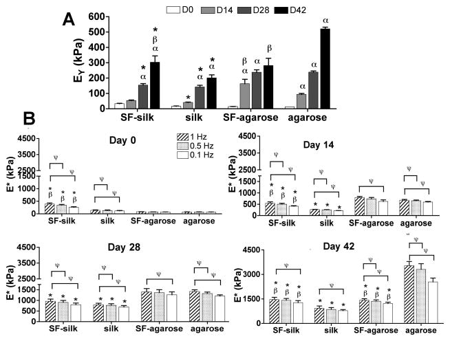
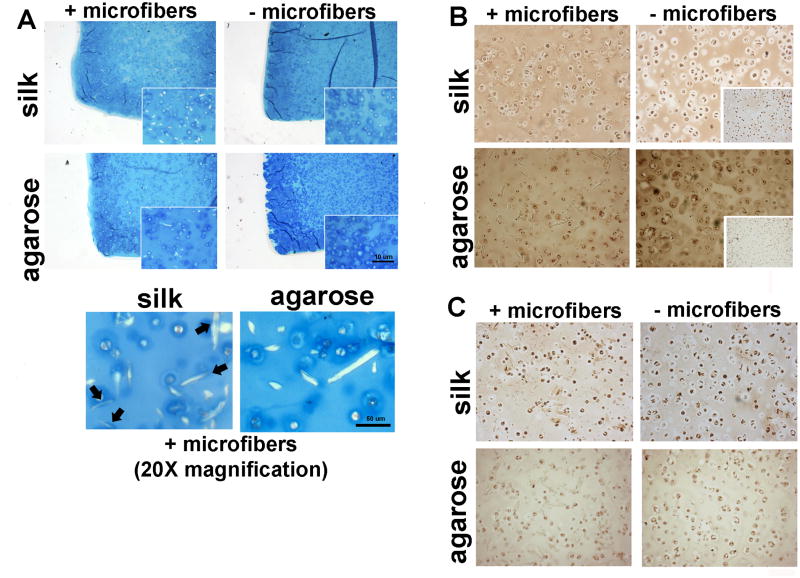
Cartilage tissue lacks an intrinsic capacity for self-regeneration due to slow matrix turnover, a limited supply of mature chondrocytes and insufficient vasculature. Although cartilage tissue engineering has achieved some success using agarose as a scaffolding material, major challenges of agarose-based cartilage repair, including non-degradability, poor tissue-scaffold integration and limited processing capability, have prompted the search for an alternative biomaterial. In this study, silk fiber-hydrogel composites (SF-silk hydrogels) made from silk microfibers and silk hydrogels were investigated for their potential use as a support material for engineered cartilage. We demonstrated the use of 100% silk-based fiber-hydrogel composite scaffolds for the development of cartilage constructs with properties comparable to those made with agarose. Cartilage constructs with an equilibrium modulus in the native tissue range were fabricated by mimicking the collagen fiber and proteoglycan composite architecture of native cartilage using biocompatible, biodegradable silk fibroin from Bombyx mori. Excellent chondrocyte response was observed on SF-silk hydrogels, and fiber reinforcement resulted in the development of more mechanically robust constructs after 42 days in culture compared to silk hydrogels alone. Thus, we demonstrate the versatility of silk fibroin as a composite scaffolding material for use in cartilage tissue repair to create functional cartilage constructs that overcome the limitations of agarose biomaterials, and provide a much-needed alternative to the agarose standard.

Keywords: Cartilage; Chondrocyte; Hydrogel; Silk; Tissue engineering.


Figures

Figure 1
Figure 1
Experimental design. Hydrogels made of silk and agarose (2% w/v and 4% w/v, respectively) were evaluated for diffusional properties using FRAP. The 4% silk and 2% agarose hydrogels exhibited similar diffusivity and were selected for further testing and reinforcement with silk microfibers. Silk microfibers of three lengths (200, 500 and >500 μm) were tested, with 500 μm microfibers producing the greatest increase in Young’s modulus in both the silk and agarose hydrogels. Cartilage tissue development was then investigated in silk and agarose hydrogels with and without the addition of the 500 μm silk microfibers.
Figure 2
Figure 2
Diffusivity of FITC–dextran in agarose and silk hydrogels. The diffusivity of FITC–dextran (MW = 70 kDa) in silk hydrogels made from varying silk protein (n = 5 for all groups). 70 kDa FITC–dextran was selected as its size is similar to the major cartilage growth factor TGF-β.
Figure 3
Figure 3
Mechanical properties of hydrogels reinforced with silk microfibers. The equilibrium and dynamic moduli of hydrogel scaffolds made of silk (A, B) and agarose (C, D) with and without incorporation of silk microfibers of different lengths (200, 500 and >500 μm) were determined in unconfined compression testing. *P < 0.05 indicates statistical significance compared with the no-fiber control group. ΨP < 0.05 indicates statistical significance compared with the 1 Hz frequency testing within the same hydrogel group, ϕP < 0.05 indicates statistical significance compared with the 500 μm hydrogel group (n = 20 for all groups).
Figure 4
Figure 4
Scanning electron micrographs of hydrogels reinforced with silk microfibers. The morphology and microarchitecture of freeze-dried, acellular (A) 4% silk hydrogel, (B) 4% SF–silk hydrogel, (C) 2% agarose hydrogel and (D) 2% SF–agarose hydrogel were visualized by SEM at 200× magnification. Silk microfibers are indicated by arrows. Scale bars are 100 μm. Insert boxes show magnified images of hydrogels. Silk microfibers are indicated by arrows.
Figure 5
Figure 5. Chondrocyte viability and biological components of constructs
Primary bovine chondrocytes were encapsulated in silk or agarose hydrogels with or without 500 μm microfibers and cultured for 42 days.(A) Live/dead assay revealed a homogeneous distribution of viable chondrocytes in all hydrogel groups at 7 and 35 days post-seeding. Silk microfibers and silk hydrogels exhibited red autofluorescence in the background. Scale bars are 200 μm. Biochemical evaluation was performed on silk and agarose hydrogels with and without 500 μm fiber reinforcement to determine total (B) DNA, (C) GAG and (D) collagen content. αP < 0.05 indicates statistical significance compared to a previous time point within the same group; βP < 0.05 indicates statistical significance compared to the same hydrogel group; *P < 0.05 indicates statistical significance compared to agarose hydrogels at the same time point (n = 5 for all groups).
Figure 6
Figure 6
Mechanical properties of engineered cartilage constructs. Constructs (n = 5 in all groups) were mechanically tested under confined compression to determine (A) the Young’s modulus (EY) and (B) the dynamic modulus (E*) every 2 weeks over the course of a 6 week culture period. αP<0.05 indicates statistical significance compared to the previous time point within the same group; βP < 0.05 indicates statistical significance compared to the same hydrogel group, * p < 0.05 indicates statistical significance compared to agarose hydrogels at the same time point; ΨP < 0.05 indicates statistical significance compared to 1 Hz frequency dynamic testing within the same group (n = 5 for all groups).
Figure 7
Figure 7
Histological evaluation of silk and agarose hydrogel cartilage constructs. (A) Alcian blue staining was used to visualize glycosaminoglycan (GAG) content within hydrogel constructs. Arrows indicate co-localization of GAGs around silk microfibers in SF–silk hydrogels, a phenomenon not observed in SF–agarose hydrogels. Immunohistochemistry of (B) collagen type II and (C) collagen type I. Collagen type II was found in a higher abundance in the no-fiber agarose control constructs compared to all other groups. Insert boxes represent negative staining.

References

    1. Murphy L, Helmick CG. The impact of osteoarthritis in the United States: a population-health perspective. Am J Nurs. 2012;112:S13. - PubMed
    1. Wang Y, Kim UJ, Blasioli DJ, Kim HJ, Kaplan DL. In vitro cartilage tissue engineering with 3D porous aqueous-derived silk scaffolds and mesenchymal stem cells. Biomaterials. 2005;26:7082. - PubMed
    1. Detterline AJ, Goldberg S, Bach BR, Jr, Cole BJ. Treatment options for articular cartilage defects of the knee. Orthop Nurs. 2005;24:361. - PubMed
    1. Korhonen RK, Laasanen MS, Toyras J, Rieppo J, Hirvonen J, Helminen HJ, Jurvelin JS. Comparison of the equilibrium response of articular cartilage in unconfined compression, confined compression and indentation. J Biomech. 2002;35:903. - PubMed
    1. Boschetti F, Pennati G, Gervaso F, Peretti GM, Dubini G. Biomechanical properties of human articular cartilage under compressive loads. Biorheology. 2004;41:159. - PubMed

Publication types