. 2015 Feb;29(1):103-12.
doi: 10.1016/j.tiv.2014.09.008. Epub 2014 Oct 2.

Validation of murine and human placental explant cultures for use in sex steroid and phase II conjugation toxicology studies

Affiliations

Validation of murine and human placental explant cultures for use in sex steroid and phase II conjugation toxicology studies

Brittany L Sato et al. Toxicol In Vitro. 2015 Feb.

Abstract

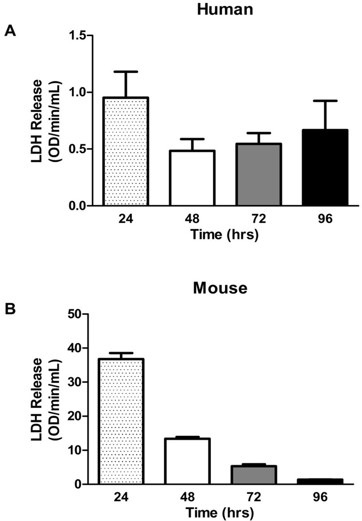
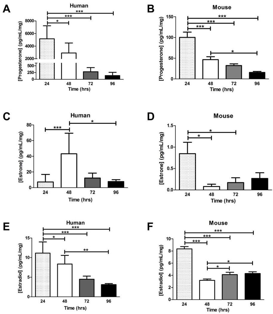
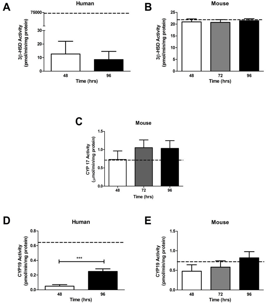
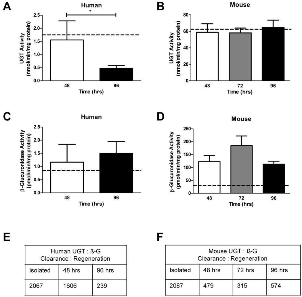
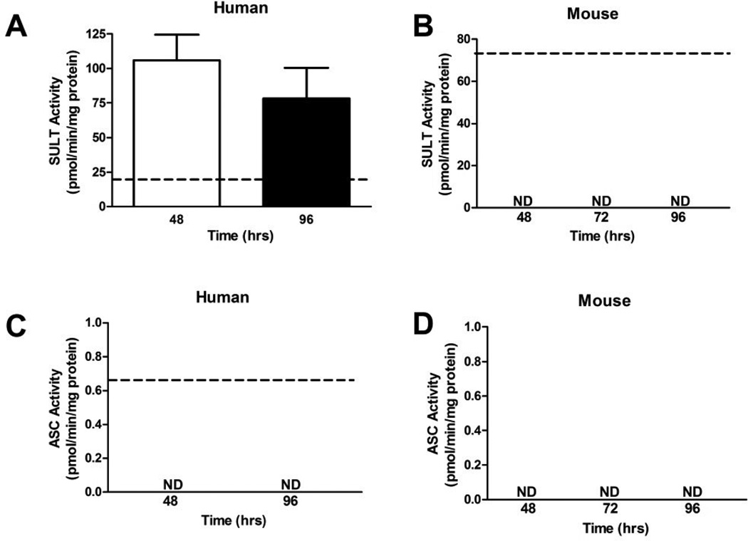
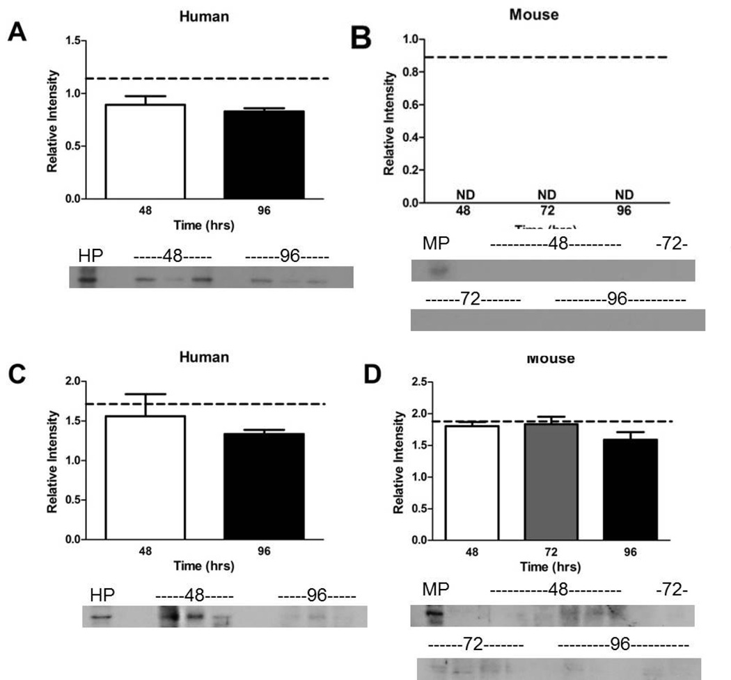
Human primary placental explant culture is well established for cytokine signaling and toxicity, but has not been validated for steroidogenic or metabolic toxicology. The technique has never been investigated in the mouse. We characterized human and mouse placental explants for up to 96 h in culture. Explant viability (Lactate dehydrogenase) and sex steroid levels were measured in media using spectrophotometry and ELISA, respectively. Expression and activities of the steroidogenic (3β-hydroxysteroid dehydrogenase, Cytochrome P45017A1, Cytochrome P45019), conjugation (UDP-glucuronosyltransferase, sulfotransferase (SULT)), and regeneration (β-glucuronidase, arylsulfatase C (ASC)) enzymes were determined biochemically in tissues with fluorimetric and spectrophotometric assays, and western blot. Explants were viable up to 96 h, but progesterone, estrone, and 17β-estradiol secretion decreased. Steroidogenic enzyme expression and activities were stable in mouse explants and similar to levels in freshly isolated tissues, but were lower in human explants than in fresh tissue (P<0.01). Human and mouse explants exhibited significantly less conjugation after 96 h, SULT was not detected in the mouse, and neither explants had active ASC, although proteins were expressed. Mouse explants may be useful for steroid biochemistry and endocrine disruption studies, but not metabolic conjugation. In contrast, human explants may be useful for studying conjugation for <48 h, but not for steroid/endocrine studies.

Keywords: Conjugation; Ex vivo culture; Mouse; Placenta; Steroidogenesis.


Figures

Figure 1
Figure 1. Viability of enriched primary explant culture over four days. LDH release is indirectly indicative of cell death
There was no significant loss of viability of A. human and B. mouse primary explants between 24 and 96 hr of culture. Bars are means ± SD for humans (n = 3 individual placentas cultured for each time point, with every placenta cultured in three wells and wells assayed in triplicate); bars are means ± SEM for mice (n = 12, 12, 11, 12 individual placentas for each time point, with every placenta cultured in three wells and wells assayed in triplicate)‥
Figure 2
Figure 2. Levels of sex steroid hormones secreted into culture medium over four days of primary explant culture
A. Progesterone levels measured in the media of human primary explants significantly decreased from 24 to 96 hr, P < 0.001, ANOVA. B. Significant decreases were observed in progesterone levels of mouse primary explants. C. Estrone levels in human primary explant media peaked at 48 hr before declining significantly, P < 0.001. D. Mouse estrone levels declined in the media after the initial 24 hr of culture, P < 0.05. E. The 17β-estradiol levels in the media of human primary explant culture displayed a significant time-dependent decrease, P < 0.001. F. The 17β-estradiol measured in mouse explant media was significantly reduced after the initial 48 hr of culture, with a slight increase at 48 and 72 hr, P < 0.001. Bars are means ± SD for human (n = 11, 12, 3, 3 placentas cultured at each time point, each well assayed in triplicate); and means ± SEM for mice (n = 12, 12, 11, 12 placentas cultured at each time point, each well assayed in triplicate). Horizontal bars indicate significance between individual groups determined with Tukey’s post hoc test. * = P < 0.05, ** = P < 0.01, *** = P < 0.001. The ANOVA results for total trend are P values reported in the legend.
Figure 3
Figure 3. The activities of steroidogenic enzymes over four days of primary explant culture
A. The activity of 3β-HSD was not significantly altered in human primary explants between 48 and 96 hr; however levels were significantly lower than 3β-HSD activity in freshly isolated human placental tissue, depicted by dotted line (n = 3 human placentas cultured in individual wells at each time point, with each well assayed in triplicate). 5). B. 3β-HSD activity in mouse placental explants did not change between 48 and 96 hr of culture, nor differ from activity levels in freshly isolated mouse placental tissue (n = 6, 6 and 7 placentas cultured in individual wells at each time point, with each well assayed in triplicate). 5). C. Levels of CYP17 activity in mouse placental explants did not significantly differ between 48 and 96 hr, nor vary from isolated mouse placental tissue (n = 6, 6, 7, individually cultured placentas, each assayed in triplicate). D. Activity of CYP19 in human placental significantly increased between 48 and 96 hr of culture and was significantly lower than activity measured in isolated placental tissue (n = 5 and 3 individual placentas cultured separately for each timepoint, then assayed in triplicate). E. CYP19 activity levels were similar in isolated mouse placental tissue and mouse placental explants cultured between 48 and 96 hr (n = 6, 6, 7 individual placentas cultured separately for each timepoint, then assayed in triplicate). The dotted line represents mean of isolated pooled placenta (n = 5 for humans and mice), bars are means ± SD for humans; bars are means ± SEM for mice. *** = P < 0.001 between time-points.
Figure 4
Figure 4. The activities of UGT (steroid clearance) and β-G (steroid regeneration) enzymes over four days of primary explant culture
A. Activity levels of UGT enzymes significantly decreased between the 48 to 96 hr in human primary explant cultures, P < 0.05. B. Mouse placental explant did not experience any alterations to UGT activity between 48 and 96 hr of culture, nor differ from levels measured in the freshly isolated mouse placental tissue. C. Levels of β-G activity were not altered between 48 and 96 hr of culture in human primary explants and were similar to freshly isolated tissue. D. Mouse placental explant β-G activity was consistent between 48 and 96 hr of culture, but at all time-points was significantly higher than activity in isolated mouse placental tissue. E. Ratios of UGT: β-G showing progressive decline of clearance by glucuronidation in human cultures. F. Ratios of UGT: β-G showing immediate loss of glucuronidation that is maintained at ~25% of fresh tissue levels during culture of mouse explants. Dotted line in A–D represents mean of isolated pooled placenta (n = 5 for humans and mice), bars are means ± SD for humans (n = 5, 3, 5); bars are means ± SEM for mice (n = 6, 5, 6, 5).
Figure 5
Figure 5. The activities of SULT (steroid clearance) and ASC (steroid regeneration) enzymes over four days of primary explant culture
A. SULT activity was similar after 48 and 96 hr of culture in human placental explants. Explants activity levels were significantly higher than levels measured in isolated human placental tissue, dotted line (n = 3, placentas cultured at each time point, then assayed in triplicate). B. SULT activity was only detectable in isolated mouse placental tissue and not in mouse placental explants. C. ASC activity was only detectable in isolated human placental tissue, but not detectable in human placental explants. D. ASC activities were not detected in fresh mouse tissue or placental explants. The dotted line represents a pool of n = 5 freshly isolated mouse or human placentas for SULT, and n = 10 for ASC, with the pool assayed once in triplicate. Enzyme activities were determined in triplicate for each sample. Bars are means ± SD.
Figure 6
Figure 6. The expression of SULT and ASC over four days of primary explant culture
A. The expression of SULT proteins in human placental explants did not change between 48 and 96 hr and was similar to expression of SULT in isolated human placental tissue. B. The expression of SULT proteins was only detectable in isolated mouse placental tissue but not in mouse explants. C. Expression of ASC proteins was similar in isolated human placental tissue and human placental explants. D. Although ASC activity was undetectable in mouse placental explants (Fig. 5D), expression of the proteins was measurable across the cultured time points and isolated mouse placental tissue. Dotted line represents mean of isolated pooled placenta (n = 5 in the pool for humans and mice, assayed once in triplicate), bars are means ± SD for humans (n = 3 individual placentas cultured, then each culture assayed in triplicate); bars are means ± SEM for mouse (n = 6, 5, 5 individual placentas cultured, then each culture assayed in triplicate). HP and MP are pooled human (n = 5) and pooled mouse (n = 5) freshly isolated placental tissue for western blot.

Similar articles

Cited by

References

    1. Ahmed N, Murphy B. The effects of various hormones on human chorionic gonadotropin production in early and late placental explant cultures. American Journal of Obstetrics and Gynecology. 1988;159:1220–1227. - PubMed
    1. Begum-Hasan J, Senterman M, Gillett P, LaPlante Branchaud C, Murphy BE. Effect of maternal serum on viability and function of early human placental explants. In Vitro Cellular and Developmental Biology - Animals. 1993;29A:505–511. - PubMed
    1. Ben-Zimra M, Koler M, Melamed-Book N, Arensburg J, Payne AH, Orly J. Uterine and placental expression of steroidogenic genes during rodent pregnancy. Molecular and Cellular Endocrinology. 2002;187:223–231. - PubMed
    1. Benyo DF, Miles TM, Conrad KP. Hypoxia Stimulates Cytokine Production by Villous Explants from the Human Placenta. Journal of Clinical Endocrinology and Metabolism. 1997;82:1582–1588. - PubMed
    1. Carter AM. Animal models of human placentation - a review. Placenta. 2007;28(Supplement):S41–S47. - PubMed

Publication types

MeSH terms

LinkOut - more resources