. 2014 Oct 6:4:6528.
doi: 10.1038/srep06528.

Assembly dynamics and the roles of FliI ATPase of the bacterial flagellar export apparatus

Affiliations

Assembly dynamics and the roles of FliI ATPase of the bacterial flagellar export apparatus

Fan Bai et al. Sci Rep. .

Abstract

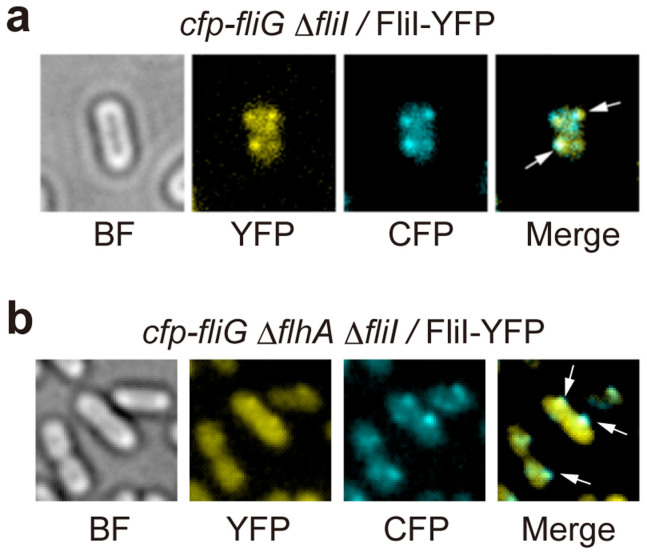
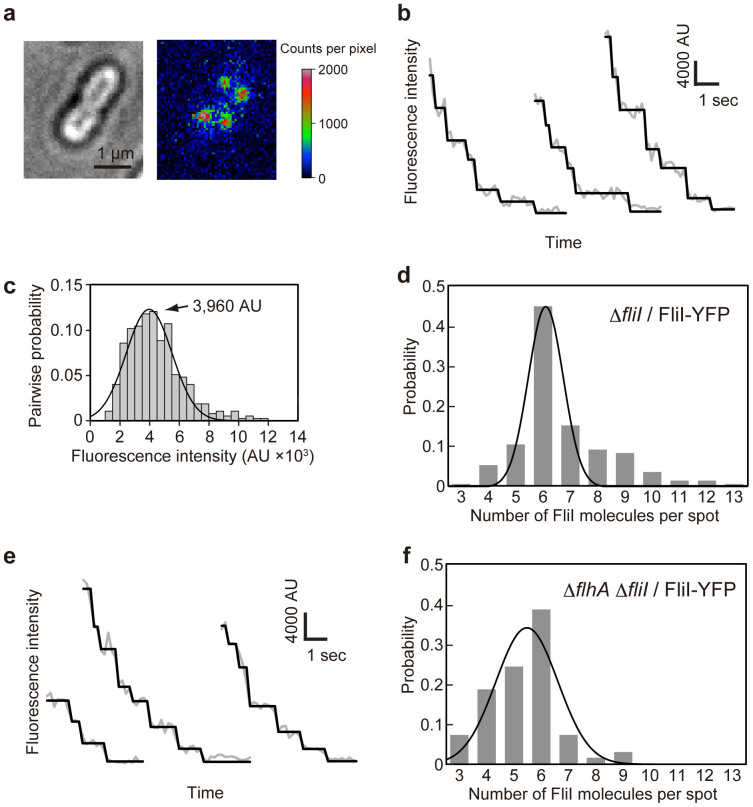
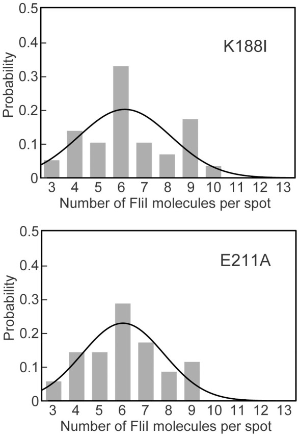
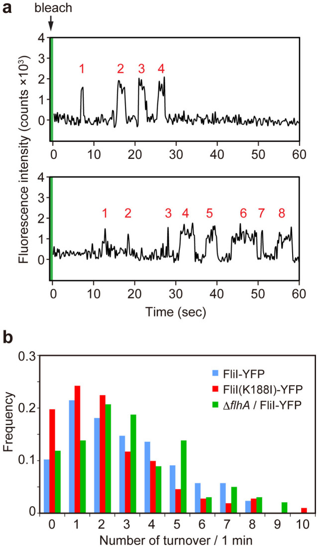
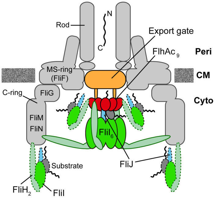
For construction of the bacterial flagellum, FliI ATPase forms the FliH2-FliI complex in the cytoplasm and localizes to the flagellar basal body (FBB) through the interaction of FliH with a C ring protein, FliN. FliI also assembles into a homo-hexamer to promote initial entry of export substrates into the export gate. The interaction of FliH with an export gate protein, FlhA, is required for stable anchoring of the FliI6 ring to the gate. Here we report the stoichiometry and assembly dynamics of FliI-YFP by fluorescence microscopy with single molecule precision. More than six FliI-YFP molecules were associated with the FBB through interactions of FliH with FliN and FlhA. Single FliI-YFP molecule exchanges between the FBB-localized and free-diffusing ones were observed several times per minute. Neither the number of FliI-YFP associated with the FBB nor FliI-YFP turnover rate were affected by catalytic mutations in FliI, indicating that ATP hydrolysis by FliI does not drive the assembly-disassembly cycle of FliI during flagellar assembly. We propose that the FliH2FliI complex and FliI6 ring function as a dynamic substrate carrier and a static substrate loader, respectively.


Figures

Figure 1
Figure 1. Localization of FliI-YFP to the FBB.
Bright field (BF) and epi-fluorescence images of FliI-YFP (YFP) and CFP-FliG (CFP) in (a) Salmonella YVM029 (cfp-fliG ΔfliI) strain transformed with pJSV203 (FliI-YFP) and (b) YVMA013 (ΔflhA ΔfliI cfp-fliG) strain carrying pJSV203. The fluorescence images of CFP-FliG (cyan) and FliI-YFP (yellow) are merged in the right panel. Arrows point to the FliI-YFP spots that co-localize with the CFP-FliG spots.
Figure 2
Figure 2. Measurement of the number of FliI-YFP molecules associated with the FBB using step photobleaching.
(a) Bright field image (left) and 2D intensity plot (right) of an epi-fluorescence image (false-color) of Salmonella MKM30 (ΔfliI) strain harboring pJSV203 (FliI-YFP). (b) Typical examples show continuous photobleaching intensity trace (gray line connecting dots) for a single FliI-YFP spot in MKM30 harboring pJSV203. Filtered intensity (black line) is overlaid on the raw intensity (gray). (c) Histogram of the pairwise difference distribution for photobleaching traces. The highest peak was fitted by a Gaussian function (black line). In total 73 FliI–YFP spots were analyzed. (d) Histogram of the estimated number of FliI-YFP molecules per spot in MKM30 carrying pJSV203. In total 230 FliI-YFP spots were analyzed. (e) and (f) Effect of flhA deletion on the number distribution of FliI-YFP molecules per spot observed in Salmonella NH0027 (ΔflhA ΔfliI) strain harboring pJSV203. (e) Three typical examples show continuous photobleaching intensity trace for a single FliI-YFP spot. (f) Histogram of the estimated number of FliI-YFP molecules per spot in NH0027 carrying pJSV203. In total 70 FliI-YFP spots were analyzed. The number distributions were fitted by Gaussian functions (black line) in (d) and (f).
Figure 3
Figure 3. Effects of K188I and E211A catalytic mutations on the number of FliI-YFP molecules per spot.
The number of photobleaching steps in each FliI(K188I)-YFP (a) and FliI(E211A)-YFP (b) spot was counted in Salmonella MKM30 (ΔfliI) strain transformed with pSY001 and pSY002, respectively. In total, 58 FliI(K188I)-YFP and 50 FliI(E211A)-YFP spots were analyzed. The number distributions were fitted by Gaussian functions (black line).
Figure 4
Figure 4. Observation of FliI-YFP turnover at the flagellar base.
(a) Typical two examples of the fluorescent intensity trace in each FliI-YFP spot in Salmonella MKM30 strain harboring pJSV203 under continuous TIRF illumination after one-shot photobleaching using a strong excitation laser (green band). Peaks labeled with numbers in red show intensity recovery of a single FliI-YFP molecule within a time period of 60 seconds. (b) The number of times of single FliI-YFP molecule turnover per minute. Each YFP spot was counted in Salmonella MKM30 (ΔfliI) strain harboring pJSV203 (as indicated as FliI-YFP, light blue) (n = 89) and pSY001 (as indicated as FliI(K188I)-YFP, red) (n = 112) and NH0027 (ΔflhA ΔfliI) carrying pJSV203 (as indicated as ΔflhA/FliI-YFP, green) (n = 102).
Figure 5
Figure 5. Schematic diagrams of the bacterial flagellar export apparatus.
The export gate made of FlhA, FlhB, FliO, FliP, FliQ and FliR are located within the central pore of the MS ring. The C-terminal cytoplasmic domain of FlhA (FlhAC) forms a nonameric ring structure and projects into the cavity of the C ring formed by FliG, FliM, and FliN. FliI forms a homo-hexamer (FliI6). The FliI6 ring firmly associates with the FBB through interactions of FliH with both FliN and FlhA. FliI also forms the FliH2-FliI complex and binds to the FBB through the FliH-FliN and FliH-FlhA interactions. During flagellar assembly, the FliH2FliI complex binds to FliJ and export substrate in the cytoplasm and acts as a dynamic carrier (dashed line) to deliver FliJ and export substrate to the C and FlhAC9 rings. Upon formation of the FliH12FliI6FliJ ring complex (continuous line) on the FlhAC9-FlhBC platform, the FliI6 ring can act as a substrate loader to promote the initial entry of the substrate into the gate. A specific interaction of FliJ located at the center of the FliI6 ring with a flexible linker of FlhA allows the export gate to translocate flagellar protein in a PMF-dependent manner.

References

    1. Chevance F. F. & Hughes K. T. Coordinating assembly of a bacterial macromolecular machine. Nat. Rev. Microbiol. 6, 455–465 (2008). - PMC - PubMed
    1. Minamino T., Imada K. & Namba K. Mechanisms of type III protein export for bacterial flagellar assembly. Mol. BioSyst. 4, 1105–1115 (2008). - PubMed
    1. Minamino T. Protein export through the bacterial flagellar type III export pathway. Biochim. Biophys. Acta. 1843, 1642–1648 (2014). - PubMed
    1. Francis N. R., Sosinsky G. E., Thomas D. & DeRosier D. J. Isolation, characterization, and structure of bacterial flagellar motors containing the switch complex. J. Mol. Biol. 235, 1261–1270 (1994). - PubMed
    1. González-Pedrajo B., Minamino T., Kihara M. & Namba K. Interactions between C ring proteins and export apparatus components: a possible mechanism for facilitating type III protein export. Mol. Microbiol. 60, 984–998 (2006). - PubMed

Publication types

MeSH terms

LinkOut - more resources