Review
. 2015 Jan;16(1):35-47.
doi: 10.1111/tra.12233. Epub 2014 Nov 5.

Role of flippases, scramblases and transfer proteins in phosphatidylserine subcellular distribution

Affiliations
Review

Role of flippases, scramblases and transfer proteins in phosphatidylserine subcellular distribution

Hannah M Hankins et al. Traffic. 2015 Jan.

Abstract

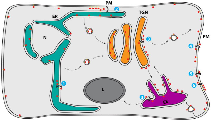
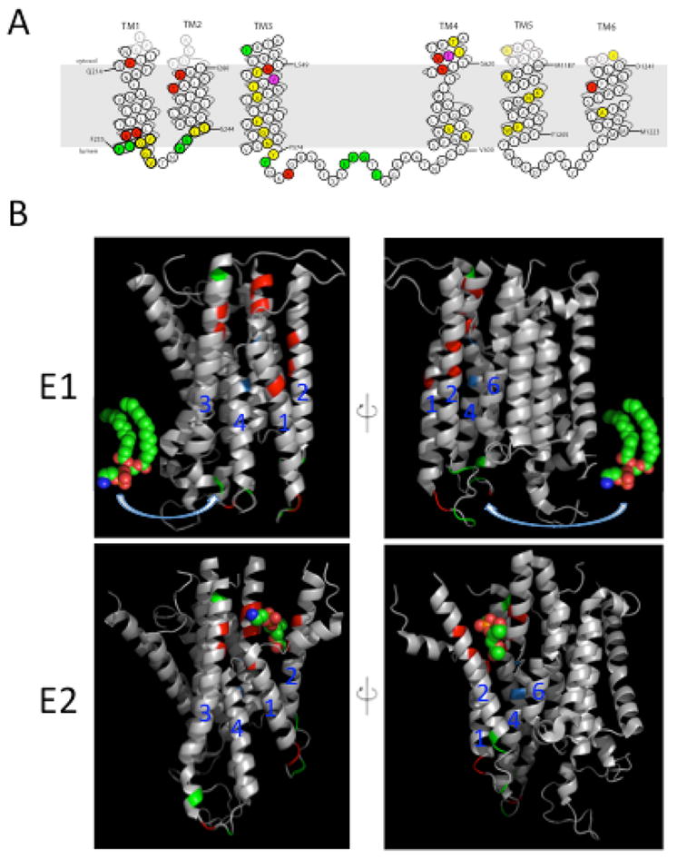
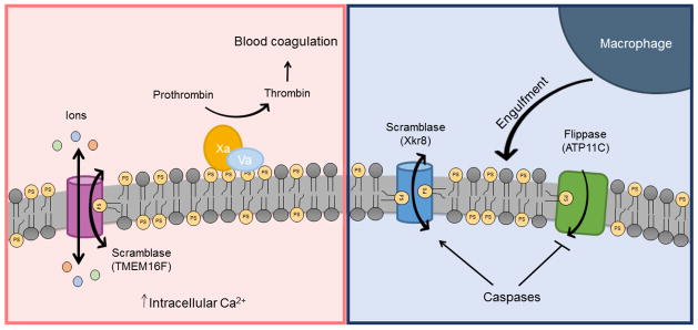
It is well known that lipids are heterogeneously distributed throughout the cell. Most lipid species are synthesized in the endoplasmic reticulum (ER) and then distributed to different cellular locations in order to create the distinct membrane compositions observed in eukaryotes. However, the mechanisms by which specific lipid species are trafficked to and maintained in specific areas of the cell are poorly understood and constitute an active area of research. Of particular interest is the distribution of phosphatidylserine (PS), an anionic lipid that is enriched in the cytosolic leaflet of the plasma membrane. PS transport occurs by both vesicular and non-vesicular routes, with members of the oxysterol-binding protein family (Osh6 and Osh7) recently implicated in the latter route. In addition, the flippase activity of P4-ATPases helps build PS membrane asymmetry by preferentially translocating PS to the cytosolic leaflet. This asymmetric PS distribution can be used as a signaling device by the regulated activation of scramblases, which rapidly expose PS on the extracellular leaflet and play important roles in blood clotting and apoptosis. This review will discuss recent advances made in the study of phospholipid flippases, scramblases and PS-specific lipid transfer proteins, as well as how these proteins contribute to subcellular PS distribution.

Keywords: P-type ATPase; flippase; lipid transfer protein; membrane asymmetry; phosphatidylserine; scramblase.


Figures

Figure 1
Figure 1
Distribution of phosphatidylserine (PS) throughout the cell. (1) PS is synthesized on the cytosolic leaflet of the endoplasmic reticulum (ER). Rapid, energy-independent flip-flop occurs in the ER membrane, but PS is preferentially localized within the lumenal leaflet, either by retention through interaction with abundant lumenal components or because (2) PS in the cytosolic leaflet is transported by non-vesicular means from the ER to plasma membrane (PM) by lipid transport proteins (LTPs). (3) PS that travels through the secretory system remains within the lumenal leaflet until it is flipped by P4-ATPases in the trans-Golgi network (TGN) and early endosomes (EE) to the cytosolic leaflet. (4) P4-ATPases also flip PS at the PM, ensuring little PS is exposed in the extracellular leaflet. (5) When activated during the process of apoptosis or blood clotting, scramblases break down the lipid asymmetry of the PM, causing exposure of PS on the outer leaflet. (6) PS exposed on the extracellular leaflet of apoptotic cells acts as an “eat me” signal for engulfment by macrophages.
Figure 2
Figure 2
Amino acid residues in the membrane domain of Dnf1 that determine substrate specificity. (A) Topology of the first 6 transmembrane (TM) segments of Dnf1 highlighting residues important for substrate specificity and transport. Dnf1 normally recognizes and transports lyso-PC and lyso-PE, but specific point mutations changing the residues highlighted in red allow recognition of PS. Mutations in residues highlighted in green perturb recognition of PC without altering translocation of PE. Changes in only two residues, highlighted in purple, alter recognition of both PC and PS. Mutation of residues in yellow reduced activity without altering substrate specificity. (B) Two potential translocation pathways for flipping phospholipid substrate. The phospholipid headgroup may engage the entry gate residues in the E1 conformation, then slide along a grooved formed by TM segments 1, 3 and 4 (left) or TM 2, 4 and 6 (right) and dock in the exit gate in the E2 conformation. The crystal structures of the Ca2+-ATPase membrane domain in the E1 (PBD ISU4) and E2 (PDB 3W5C) conformational states are shown with the positions homologous to substrate-defining residues of the P4-ATPases highlighted. Red and green positions represent the Dnf1 residues shown in (A) involved in PS and PC recognition, respectively. Positions highlighted in blue represent the P+1 Ile and N359 in ATP8A2. The PE molecule in the E1 images derived from PDB 3B74 while the PE headgroup in the E2 images co-crystallized with the Ca2+-ATPase.
Figure 3
Figure 3
Two distinct pathways for phospholipid scrambling during blood coagulation and apoptosis. During the process of blood coagulation, an elevation in intracellular Ca2+ level stimulates scramblase activity on the PM of platelets (left). The scramblase functions as both a non-selective ion channel and phospholipid scramblase with two distinct pathways for ion and phospholipid transport. Phospholipid scrambling causes exposure of PS on the surface where it acts as a platform on which coagulation protein complexes assemble. During apoptosis, caspases both activate scramblase activity, proposed to be mediated by Xkr8, and inactivate flippase activity (Atp11C) by cleaving at their caspase-recognition sites (right). Phospholipid scrambling causes PS to be exposed on the surface of the apoptotic cell where it acts as an “eat me” signal for macrophages.

References

    1. Van Meer G, Voelker DR, Feigenson GW. Membrane lipids: where they are and how they behave. Nat Rev Mol Cell Biol. 2008 Feb;9(2):112–24. - PMC - PubMed
    1. Bell RM, Ballas LM, Coleman RA. Lipid topogenesis. J Lipid Res. 1981 Mar 1;22(3):391–403. - PubMed
    1. Zinser E, Sperka-Gottlieb CD, Fasch EV, Kohlwein SD, Paltauf F, Daum G. Phospholipid synthesis and lipid composition of subcellular membranes in the unicellular eukaryote Saccharomyces cerevisiae. J Bacteriol. 1991 Mar 1;173(6):2026–34. - PMC - PubMed
    1. Zinser E, Daum G. Isolation and biochemical characterization of organelles from the yeast, Saccharomyces cerevisiae. Yeast. 1995 May 1;11(6):493–536. - PubMed
    1. Bretscher MS. Membrane Structure: Some General Principles. Science. 1973 Aug 17;181(4100):622–9. - PubMed

Publication types

MeSH terms

LinkOut - more resources