. 2014 Nov;128(5):651-63.
doi: 10.1007/s00401-014-1345-4. Epub 2014 Oct 5.

Inflammatory dysregulation of blood monocytes in Parkinson's disease patients

Affiliations

Inflammatory dysregulation of blood monocytes in Parkinson's disease patients

Veselin Grozdanov et al. Acta Neuropathol. 2014 Nov.

Abstract

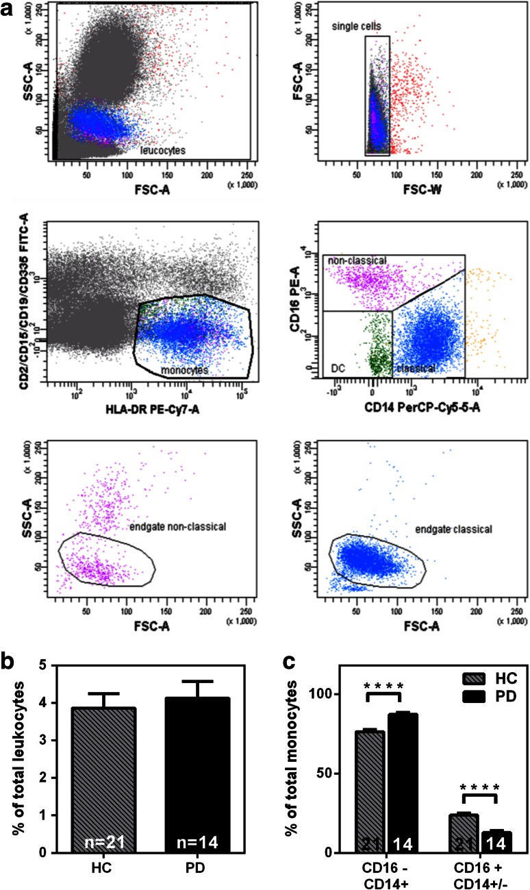
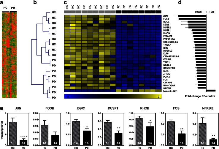
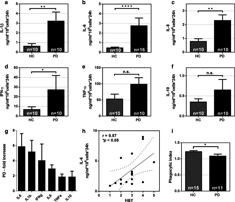
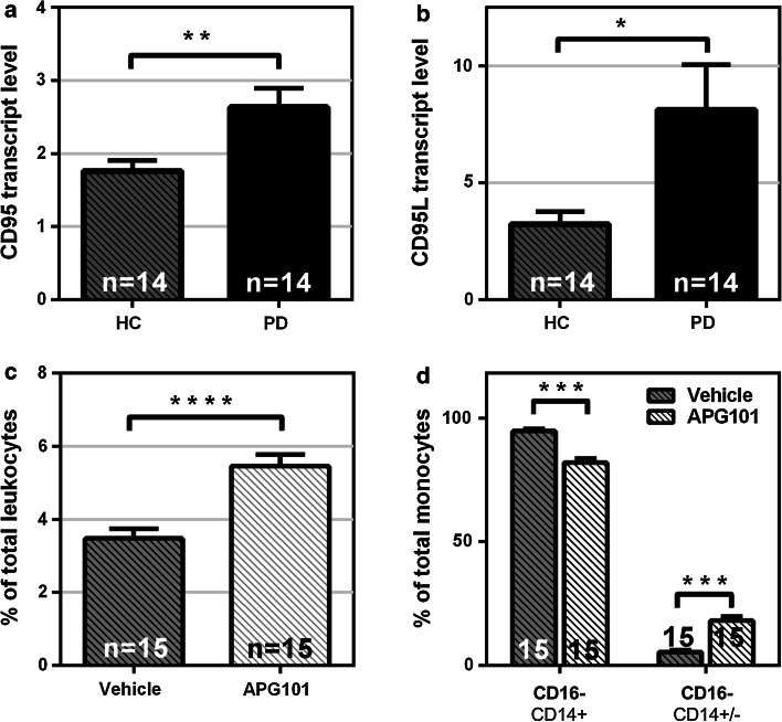
Despite extensive effort on studying inflammatory processes in the CNS of Parkinson's disease (PD) patients, implications of peripheral monocytes are still poorly understood. Here, we set out to obtain a comprehensive picture of circulating myeloid cells in PD patients. We applied a human primary monocyte culture system and flow cytometry-based techniques to determine the state of monocytes from PD patients during disease. We found that the classical monocytes are enriched in the blood of PD patients along with an increase in the monocyte-recruiting chemoattractant protein CCL2. Moreover, we found that monocytes from PD patients display a pathological hyperactivity in response to LPS stimulation that correlates with disease severity. Inflammatory pre-conditioning was also reflected on the transcriptome in PD monocytes using next-generation sequencing. Further, we identified the CD95/CD95L as a key regulator for the PD-associated alteration of circulating monocytes. Pharmacological neutralization of CD95L reverses the dysregulation of monocytic subpopulations in favor of non-classical monocytes. Our results suggest that PD monocytes are in an inflammatory predisposition responding with hyperactivation to a "second hit". These results provide the first direct evidence that circulating human peripheral blood monocytes are altered in terms of their function and composition in PD patients. This study provides insights into monocyte biology in PD and establishes a basis for future studies on peripheral inflammation.


Figures

Fig. 1
Fig. 1
Classical (CD14+CD16−) monocytes are enriched in PD patients. a Representative FACS analysis of monocyte subpopulations from peripheral blood. Monocytes are gated out of other immune cells (FITC—CD2/CD15/CD19/CD335) by HLA-DR staining (PE-Cy7) and monocyte subpopulations characterized based on CD14 (PerCP-Cy5.5) and CD16 (PE) staining. b Total number of monocytes in peripheral blood as a percentage of total leukocytes. No significant difference between PD patients’ and healthy controls’ blood was found (Healthy controls n = 21, PD n = 14). c CD14+CD16− (classical) monocytes are enriched and CD14−/+CD16+ monocytes are decreased in the blood of PD patients. (Healthy controls n = 21, PD patients n = 14). Bars mean ± SEM, ****p < 0.0001
Fig. 2
Fig. 2
Transcriptome-wide analysis of gene expression in PD and healthy control monocytes by next-generation sequencing reveals a distinct expression signature in PD monocytes. a Heatmap of 8,862 genes expressed over the threshold (FPKM >5). b Unsupervised hierarchical clustering based on the expression of 8,862 genes in blood monocytes successfully identified most PD cases and clustered them away from healthy controls. Clustering by complete linkage and cosine correlation. c Differential expression of selected top genes in PD monocytes. d Fold change of expression in PD monocytes vs. healthy controls’ monocytes. e Validation of selected genes’ expression in an independent cohort (healthy controls n = 13, PD patients n = 14) by qRT-PCR. Bars represent mean ± SEM, *p < 0.05, **p < 0.01, ****p < 0.0001
Fig. 3
Fig. 3
PD monocytes are hyperactive upon LPS stimulation. Monocytes from PD patients secrete significantly higher levels of IL-1β, IL-6, IL-8 and IFNγ when stimulated with 1 ng/ml LPS compared to monocytes from age-matched healthy controls (a–d). No significant difference in cytokine secretion was observed for TNFα (p = 0.05), and IL-10 (p = 0.52) (e, f). Bars represent mean ± SEM. PD patients (n = 10), healthy controls (n = 10), *p < 0.05, **p < 0.01, *** p < 0.001, ****p < 0.0001. g Fold increase in cytokine production of monocytes from PD patients vs. healthy controls. h Correlation between IL-6 release after 24 h stimulation with 1 ng/ml LPS and PD severity staged with the H&Y scale. Dotted lines indicate the 95 % CI of the linear regression. r Spearman’s correlation coefficient. i Decreased phagocytic function of Parkinson’s disease monocytes, healthy controls (n = 15), PD patients (n = 11). Bars represent mean ± SEM, *p < 0.05
Fig. 4
Fig. 4
The CCR2–CCL2 axis is activated in PD. a Monocytes from PD patients release significantly higher levels of CCL2 upon LPS stimulation (LPS 1 ng/ml, healthy controls n = 10, Parkinson’s disease patients n = 10, *p < 0.05). b CCL2 is significantly increased (*p < 0.05) in the serum of PD patients (n = 53) when compared to healthy controls (n = 34). Bars represent mean ± SEM
Fig. 5
Fig. 5
FAS/FASLG upregulation in monocytes from PD patients. The expression of both FAS (a) and FASLG (b) is upregulated in unstimulated PD monocytes (Healthy controls n = 14, PD patients n = 14). c Monocytes are enriched in PD leukocyte cultures after 48 h treatment with FASLG-inhibitor APG101 (n = 15, APG 0.25 mg/ml). d Classical (CD14+CD16−) monocytes are decreased and non-classical (CD14−CD16+) monocytes are increased by treatment of PD leukocytes with APG101 in culture (n = 15). Bars represent mean ± SEM. *p < 0.05, **p < 0.01, ***p < 0.001, ****p < 0.0001

References

    1. Ahmed I, Tamouza R, Delord M, Krishnamoorthy R, Tzourio C, Mulot C, Nacfer M, Lambert JC, Beaune P, Laurent-Puig P, Loriot MA, Charron D, Elbaz A. Association between Parkinson’s disease and the HLA-DRB1 locus. Mov Disord. 2012;27(9):1104–1110. doi: 10.1002/mds.25035. - DOI - PubMed
    1. Ajami B, Bennett JL, Krieger C, McNagny KM, Rossi FM. Infiltrating monocytes trigger EAE progression, but do not contribute to the resident microglia pool. Nat Neurosci. 2011;14:1142–1149. doi: 10.1038/nn.2887. - DOI - PubMed
    1. Appel SH, Beers DR, Henkel JS. T cell-microglial dialogue in Parkinson’s disease and amyotrophic lateral sclerosis: are we listening? Trends Immunol. 2010;31:7–17. doi: 10.1016/j.it.2009.09.003. - DOI - PMC - PubMed
    1. Auffray C, Sieweke MH, Geissmann F. Blood monocytes: development, heterogeneity, and relationship with dendritic cells. Annu Rev Immunol. 2009;27:669–692. doi: 10.1146/annurev.immunol.021908.132557. - DOI - PubMed
    1. Beier CP, Schulz JB. CD95/Fas in the brain–not just a killer. Cell Stem Cell. 2009;5:128–130. doi: 10.1016/j.stem.2009.07.008. - DOI - PubMed

Publication types

MeSH terms