. 2015 Jul 23;34(30):3968-76.
doi: 10.1038/onc.2014.328. Epub 2014 Oct 6.

P-REX1 creates a positive feedback loop to activate growth factor receptor, PI3K/AKT and MEK/ERK signaling in breast cancer

Affiliations

P-REX1 creates a positive feedback loop to activate growth factor receptor, PI3K/AKT and MEK/ERK signaling in breast cancer

L M Dillon et al. Oncogene. .

Abstract

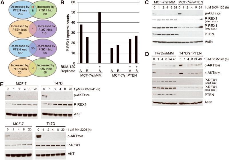
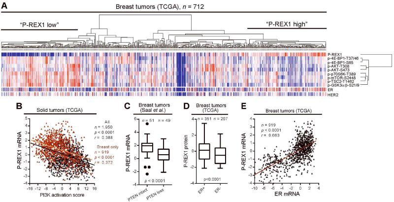
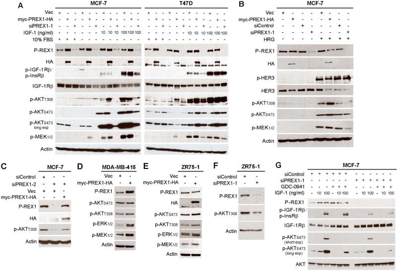
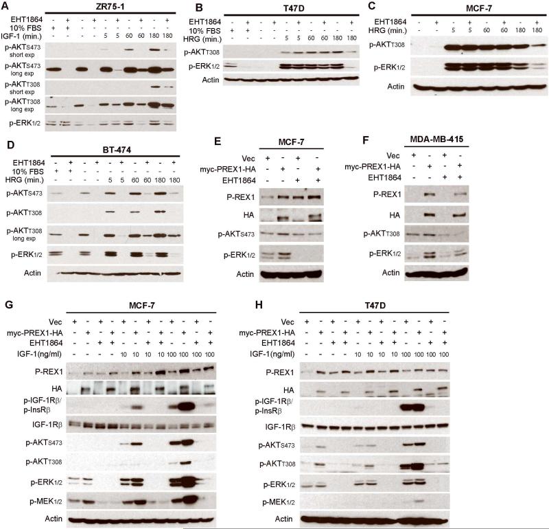
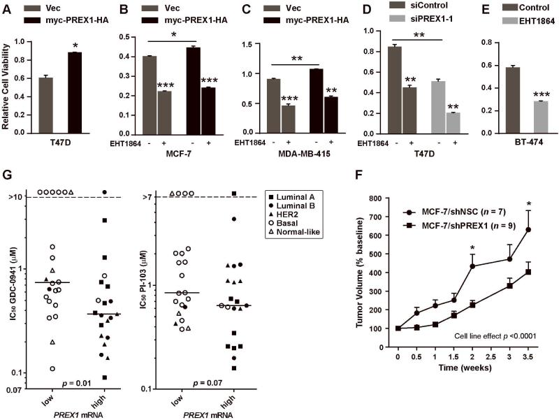
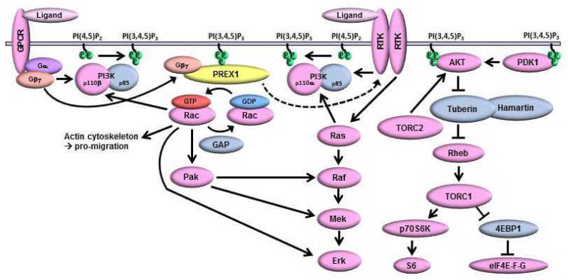
Phosphatidylinositol 3-kinase (PI3K) promotes cancer cell survival, migration, growth and proliferation by generating phosphatidylinositol 3,4,5-trisphosphate (PIP3) in the inner leaflet of the plasma membrane. PIP3 recruits pleckstrin homology domain-containing proteins to the membrane to activate oncogenic signaling cascades. Anticancer therapeutics targeting the PI3K/AKT/mTOR (mammalian target of rapamycin) pathway are in clinical development. In a mass spectrometric screen to identify PIP3-regulated proteins in breast cancer cells, levels of the Rac activator PIP3-dependent Rac exchange factor-1 (P-REX1) increased in response to PI3K inhibition, and decreased upon loss of the PI3K antagonist phosphatase and tensin homolog (PTEN). P-REX1 mRNA and protein levels were positively correlated with ER expression, and inversely correlated with PI3K pathway activation in breast tumors as assessed by gene expression and phosphoproteomic analyses. P-REX1 increased activation of Rac1, PI3K/AKT and MEK/ERK signaling in a PTEN-independent manner, and promoted cell and tumor viability. Loss of P-REX1 or inhibition of Rac suppressed PI3K/AKT and MEK/ERK, and decreased viability. P-REX1 also promoted insulin-like growth factor-1 receptor activation, suggesting that P-REX1 provides positive feedback to activators upstream of PI3K. In support of a model where PIP3-driven P-REX1 promotes both PI3K/AKT and MEK/ERK signaling, high levels of P-REX1 mRNA (but not phospho-AKT or a transcriptomic signature of PI3K activation) were predictive of sensitivity to PI3K inhibitors among breast cancer cell lines. P-REX1 expression was highest in estrogen receptor-positive breast tumors compared with many other cancer subtypes, suggesting that neutralizing the P-REX1/Rac axis may provide a novel therapeutic approach to selectively abrogate oncogenic signaling in breast cancer cells.


Figures

Figure 1
Figure 1. P-REX1 levels decrease upon PTEN loss and increase with PI3K inhibition
A) Summary of results from mass spectrometry-based proteomic screen in MCF-7 cells (red asterisk indicates P-REX1). B) P-REX1 spectral counts from mass spectrometry shown for each of duplicate runs (run B for MCF-7/shMM + BKM-120 sample failed and was excluded from analysis). C-E) Immunoblots of lysates from cells treated with inhibitors were probed with the indicated antibodies.
Figure 2
Figure 2. P-REX1 levels are inversely correlated with PI3K activation, and positively correlated with ER expression in human tumors
A) Hierarchical clustering of signal values of P-REX1 and markers of PI3K pathway activation quantified by RPPA of 712 breast tumors. Levels of ER and HER2 are shown below, but were excluded from clustering. Red = high; blue = low. B) Correlation of P-REX1 mRNA levels and PI3K activation score in all tumor types (black) and breast tumors (red) from TCGA. Pearson correlation coefficients (r) and p-values are indicated. C) Tukey box plot comparing P-REX1 mRNA levels in breast tumors stratified based on expression of an mRNA signature of PTEN-loss vs. -intact (determined in ref. [17]) by Student’s t-test. D) Comparison of P-REX1 protein levels [from tumors in (A)] in ER+ vs. ER− breast tumors as in (C) (clinical ER status determined by IHC). E) Comparison of P-REX1 and ER mRNA levels in breast tumors from TCGA as in (B).
Figure 3
Figure 3. P-REX1 activates IGF-1R/InsR, PI3K/AKT, and MEK/ERK signaling in breast cancer cells
Immunoblot analysis of lysates from cells transfected with vectors encoding myc-PREX1-HA, vector (vec) control, siRNA against P-REX1, or siControl. Panels A-C and E-G describe transiently transfected cells. Panel D describes stably transfected cells. Cells were maintained in 10% FBS (A,C-F), or serum-starved for 24 hours then stimulated ± 10-100 ng/ml IGF-1 for 10 min. (A,G), or 10 ng/ml heregulin (HRG) for 5 min. (B). In panel G, cells were pretreated ± 1 μM GDC-0941 for 2 h, then stimulated ± IGF-1.
Figure 4
Figure 4. Rac promotes activation of PI3K/AKT and MEK/ERK and is required for P-REX1-induced activation of these pathways in breast cancer
A-D) Immunoblot analysis of lysates from cells treated as indicated ± serum-starvation for 24 h, then pretreated ± 50 μM EHT1864 × 2 h, and stimulated ± 20 ng/mL HRG or 100 ng/mL IGF-1. E-H) Immunoblot analysis of lysates from cells transiently (E,G,H) or stably (F) transfected with myc-PREX1-HA or vector (vec) control, maintained in 10% FBS (E-F) or serum-starved for 24 hours (G-H), pretreated ± 50 μM EHT1864 × 2 h (E-H), then stimulated ± IGF-1 for 10 min. (G-H).
Figure 5
Figure 5. P-REX1 increases viability of breast cancer cells
A-E) Cells transiently (A-B) or stably (C) transfected with vectors encoding myc-PREX1-HA or vector (vec) control, transiently transfected with siRNA (D), or untransfected (E), were evaluated by XTT assay. In (B-E), cells were treated ± 50 μM EHT1864 for 3 days prior to assay. Mean of 3-4 replicates ± SEM is shown. *p<0.05, **p<0.01 by Student’s t-test vs. control. F) Mice were subcutaneously implanted with MCF-7/shPREX1 or MCF-7/shNSC cells. Once tumor volumes reached ~150 mm3, tumors were measured twice weekly. Data are presented as % baseline volume (mean + SEM). Data were analyzed by two-way ANOVA followed by Bonferonni post-hoc comparison between cell lines at each time point (*p<0.05; **p<0.01).G) Forty-three breast cancer cell lines were dichotomized based on P-REX1 mRNA levels, and IC50 values were compared by Mann-Whitney U-test. Legend indicates molecular subtypes. Horizontal bars indicate median values.
Figure 6
Figure 6. Model of PI3K-dependent P-REX1 positive feedback to activate Rac, IGF-1R/InsR, PI3K/AKT, and MEK/ERK signaling
Growth factor receptor tyrosine kinases (RTKs) and G-protein coupled receptors (GPCRs) activate PI3K, which produces PIP3. P-REX1 is recruited to the membrane via PH domain binding to PIP3. Activation of GPCRs induces dissociation of Gβγ subunits, which bind the P-REX1 DH domain to promote its GEF activity and Rac activation. Rac activates PI3K/p110β to increase PIP3 production, stimulates Raf/MEK signaling via Pak, interacts with p-ERK, and promotes actin cytoskeleton remodeling. P-REX1 also promotes PI3K-independent activation of IGF-1R/InsR, which can lead to further activation of PI3K and MEK signaling.

References

    1. Miller TW, Rexer BN, Garrett JT, Arteaga CL. Mutations in the phosphatidylinositol 3-kinase pathway: role in tumor progression and therapeutic implications in breast cancer. Breast Cancer Res. 2011;13(6):224. - PMC - PubMed
    1. Engelman JA. Targeting PI3K signalling in cancer: opportunities, challenges and limitations. Nat Rev Cancer. 2009;9(8):550–62. - PubMed
    1. Whitman M, Downes CP, Keeler M, Keller T, Cantley L. Type I phosphatidylinositol kinase makes a novel inositol phospholipid, phosphatidylinositol-3-phosphate. Nature. 1988;332(6165):644–6. - PubMed
    1. Maehama T, Dixon JE. The tumor suppressor, PTEN/MMAC1, dephosphorylates the lipid second messenger, phosphatidylinositol 3,4,5-trisphosphate. Journal of Biological Chemistry. 1998;273(22):13375–13378. - PubMed
    1. Maira SM, Pecchi S, Huang A, Burger M, Knapp M, Sterker D, et al. Identification and characterization of NVP-BKM120, an orally available pan-class I PI3-kinase inhibitor. Mol Cancer Ther. 2012;11(2):317–28. - PubMed

Publication types

Substances