

Femtosecond laser versus mechanical microkeratome-assisted flap creation for LASIK: a prospective, randomized, paired-eye study
- PMID: 25284975
- PMCID: PMC4181739
- DOI: 10.2147/OPTH.S68124
Femtosecond laser versus mechanical microkeratome-assisted flap creation for LASIK: a prospective, randomized, paired-eye study
Abstract
Purpose: To compare a femtosecond laser with a microkeratome for flap creation during laser in situ keratomileusis (LASIK) in terms of flap thickness predictability and visual outcomes.
Patients and methods: This was a prospective, randomized, masked, paired-eye study. Forty-four patients (34 females) who received bilateral LASIK were included. Patients were stratified by ocular dominance, and they then underwent randomization of flap creation using the femtosecond laser on one eye and undergoing the microkeratome procedure on the other one. The visual outcome differences between the corrected distance visual acuity (CDVA) at baseline and the uncorrected distance visual acuity (UDVA) on the first day postoperatively were set as the efficiency index for both groups. All visual acuity outcome results and the deviation of flap thickness were evaluated. P-values <0.05 were considered statistically significant.
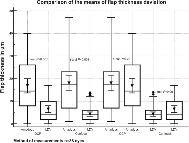
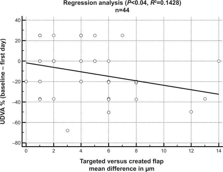
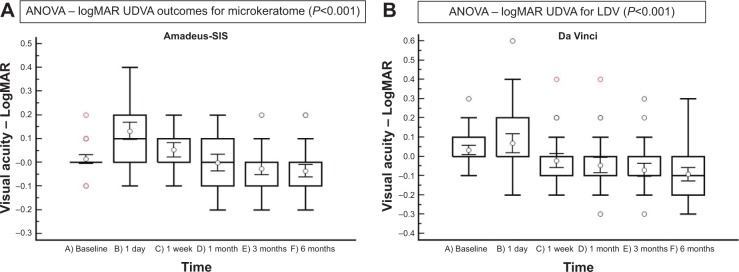
Results: The index of efficiency regarding the postoperative visual outcomes in the microkeratome group was lower (P<0.0001). This result was correlated with the difference between intended and achieved flap thickness (P=0.038; r=0.28), and a negative relationship in the regression analysis was confirmed (P<0.04; R (2)=0.1428). The UDVA in the microkeratome group improved significantly by the end of the first month (P<0.0271) in comparison to the baseline CDVA. The deviation between intended and postoperative flap thickness using either optical coherence pachymetry or Heidelberg Retinal Tomography II confocal microscopy was statistically significant (paired t-test; P<0.001) between the groups. The flap thickness deviation in the microkeratome group was higher. In the femtosecond laser group, the efficiency index was stable postoperatively (P=0.64) The UDVA improved significantly by the end of the first postoperative week (P=0.0043) in comparison to the baseline CDVA. Six months after surgery, improvement in the UDVA was significant in both groups (all P<0.001; one way analysis of variance).
Conclusion: Femtosecond laser was superior to microkeratome-assisted LASIK in terms of flap thickness predictability and the speed of visual acuity recovery. A negative relationship in the regression analysis between increasing flap thickness deviation and visual acuity recovery was confirmed.
Keywords: LASIK; femtosecond laser; flap predictability; microkeratome.
Figures
References
-
- Wirthlin AC. The ultrastable and compact Femto LDV. Cataract and Refractive Surgery Today Europe. 2007;2(3):65–66.
-
- Durrie DS, Kezirian GM. Femtosecond laser versus mechanical keratome flaps in wavefront-guided laser in situ keratomileusis: prospective contralateral eye study. J Cataract Refract Surg. 2005;31(1):120–126. - PubMed
-
- Binder PS. One thousand consecutive IntraLase laser in situ keratomileusis flaps. J Cataract Refract Surg. 2006;32(6):962–969. - PubMed
-
- Kezirian GM, Stonecipher KG. Comparison of the IntraLase femtosecond laser and mechanical keratomes for laser in situ keratomileusis. J Cataract Refract Surg. 2004;30(4):804–811. - PubMed
-
- Tran DB, Sarayba MA, Bor Z, et al. Randomized prospective clinical study comparing induced aberrations with IntraLase and Hansatome flap creation in fellow eyes: potential impact on wavefront-guided laser in situ keratomileusis. J Cataract Refract Surg. 2005;31(1):97–105. - PubMed
LinkOut - more resources
Full Text Sources
Other Literature Sources
Medical
