Review
. 2013 Jun;4(3):233-57.
doi: 10.1515/bmc-2012-0042.

Sugar glues for broken neurons

Affiliations
Review

Sugar glues for broken neurons

Vimal P Swarup et al. Biomol Concepts. 2013 Jun.

Abstract

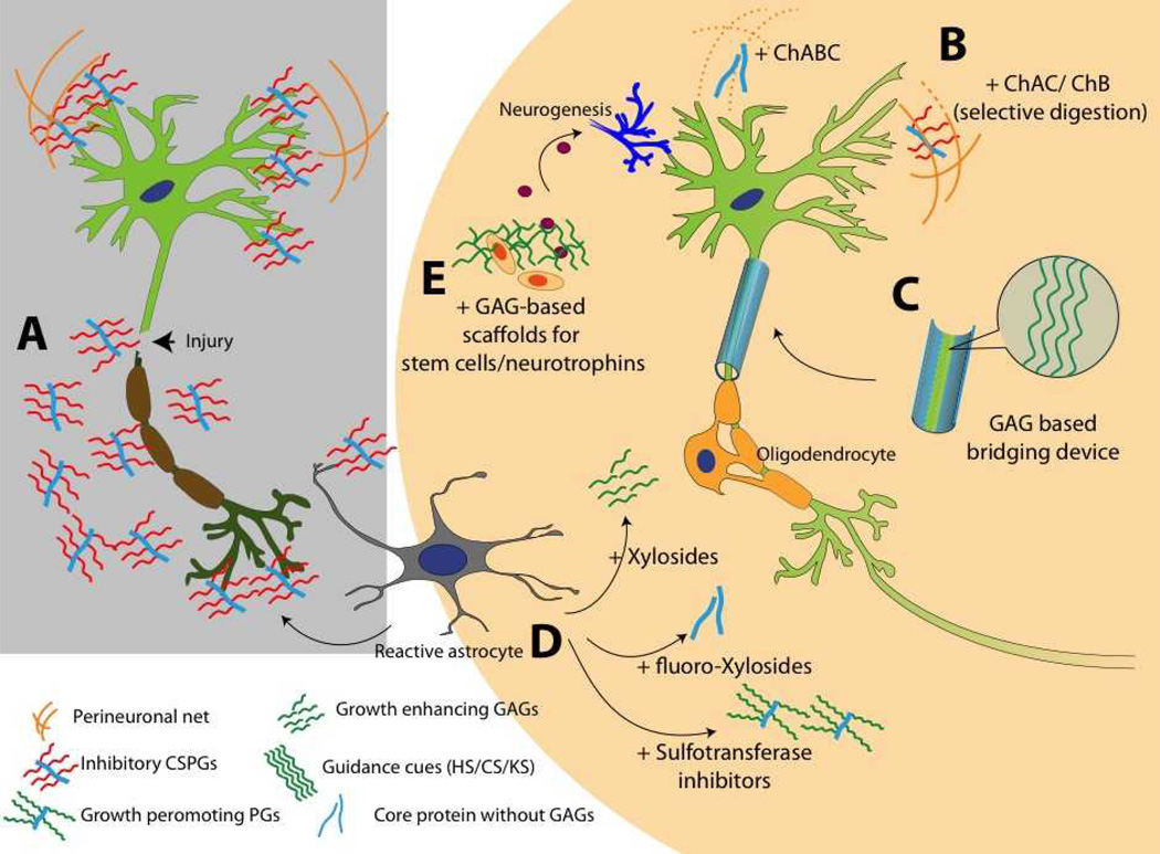
Proteoglycans (PGs) regulate diverse functions in the central nervous system (CNS) by interacting with a number of growth factors, matrix proteins, and cell surface molecules. Heparan sulfate (HS) and chondroitin sulfate (CS) are two major glycosaminoglycans present in the PGs of the CNS. The functionality of these PGs is to a large extent dictated by the fine sulfation patterns present on their glycosaminoglycan (GAG) chains. In the past 15 years, there has been a significant expansion in our knowledge on the role of HS and CS chains in various neurological processes, such as neuronal growth, regeneration, plasticity, and pathfinding. However, defining the relation between distinct sulfation patterns of the GAGs and their functionality has thus far been difficult. With the emergence of novel tools for the synthesis of defined GAG structures, and techniques for their characterization, we are now in a better position to explore the structure-function relation of GAGs in the context of their sulfation patterns. In this review, we discuss the importance of GAGs on CNS development, injury, and disorders with an emphasis on their sulfation patterns. Finally, we outline several GAG-based therapeutic strategies to exploit GAG chains for ameliorating various CNS disorders.


Figures

Figure 1
Figure 1
Schematic representation of disaccharide units found in HS, HA, KS and CS.
Figure 2
Figure 2
Receptors of HSPGs. Prominent interactions in the CNS that are mediated by HSPGs are shown. Robo receptors bind to Slit ligands, UNC-5 and DCC (deleted in colorectal cencer) receptors bind to Netrin, FGF binds to FGFR and ephrin receptors (Eph) bind to ephrin ligands. Box contains the definition for conserved protein domains. Ig: immunoglobulin; EGF, epidermal growth factor; FN3, fibronectin type III domain; TK, tyrosine kinase domain. [The figure has been modified from the review by Lee et al. (3)]
Figure 3
Figure 3
Intracellular signaling mechanism triggered by CSPG present in the glial scar. CSPG receptors are thought to be present in axons although their molecular identity is not well established. Recent studies have shown that CSPG can interact with leukocyte common antigen-related phosphatase, Nogo, or EGF receptors and lead to growth inhibition.(–67) RhoA activation eventually leads to actin depolymerization and the growth cone retraction. RhoA activation has also been shown to be associated with PKC pathway and epidermal growth factor receptor phosphorylation (EGFR) in calcium dependent manner.(67, 71) However, the process of calcium influx leading to PKC activation or EGFR activation is not well defined. Dashed arrows suggest that mediators of the represented process are yet to be identified.
Figure 4
Figure 4
Observed and potential associations of PGs and GAGs with neurological diseases in various regions of the human brain. This diagram represents a coronal section of the human brain and highlights evidence reported about the involvement of GAGs in various CNS disorders. Observations made in non-human brain samples may suggest that such effects persist in the human brain as well.
Figure 5
Figure 5
Use of sugar glues for broken neurons and GAG based therapies for injured CNS. A. Neuronal injury results in overexpression of inhibitory CSPGs from reactive astrocytes. B. Degrading CS chains to stimulate neuronal growth. ChABC is widely studied for this application; however, unspecific digestion of GAGs can damage growth promoting motifs and other important structures such as PNN. Selective digestion can be done using ChAC and ChB enzymes. C. Designing GAG based bridging device to guide regenerating axons. Surface presented HS, CS, DS, KS, or HA domains can be used to direct axonal pathfinding. D. Using small molecules to modulate GAG production at scar site. Xylosides would change composition or sulfation pattern of the GAGs secreted from reactive astrocytes. Addition of fluoro-xylosides would inhibit GAG biosynthesis and result in generation of core protein without inhibitory GAG chains. Sulfotransferase inhibitors can be used to stop production of inhibitory sulfation patterns present in CSPGs released from reactive astrocytes. E. Bioactive scaffolds can be designed by using GAGs such as HA, HS, CS, DS or KS. Such scaffolds can be used to deliver stem cells or neurotropic factors at the scar site. A combination of two or more such approaches can be utilized to create effective therapies for neurological disorders.

References

    1. Bao X, Mikami T, Yamada S, Faissner A, Muramatsu T, Sugahara K. Heparin-binding growth factor, pleiotrophin, mediates neuritogenic activity of embryonic pig brain-derived chondroitin sulfate/dermatan sulfate hybrid chains. The Journal of Biological Chemistry. 2005;280(10):9180–9191. - PubMed
    1. Bülow HE, Hobert O. Differential sulfations and epimerization define heparan sulfate specificity in nervous system development. Neuron. 2004;41(5):723–736. - PubMed
    1. Lee J, Chien C. When sugars guide axons: insights from heparan sulphate proteoglycan mutants. Nature Reviews Genetics. 2004;5(12):923–935. - PubMed
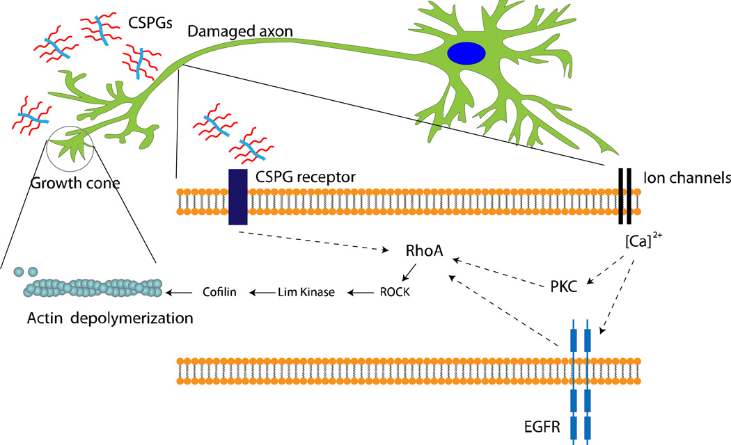
    1. Asher RA, Morgenstern DA, Moon LDF, Fawcett JW. Chondroitin sulphate proteoglycans: inhibitory components of the glial scar. Progress in Brain Research. 2001;132:611–619. - PubMed
    1. Snow DM, Lemmon V, Carrino DA, Caplan AI, Silver J. Sulfated proteoglycans in astroglial barriers inhibit neurite outgrowth in vitro. Experimental Neurology. 1990;109(1):111–130. - PubMed

Publication types

MeSH terms

LinkOut - more resources