. 2014 Dec:77:11-9.
doi: 10.1016/j.yjmcc.2014.09.028. Epub 2014 Oct 5.

Role of the ubiquitin-proteasome system in cardiac dysfunction of adipose triglyceride lipase-deficient mice

Affiliations

Role of the ubiquitin-proteasome system in cardiac dysfunction of adipose triglyceride lipase-deficient mice

Marion Mussbacher et al. J Mol Cell Cardiol. 2014 Dec.

Abstract

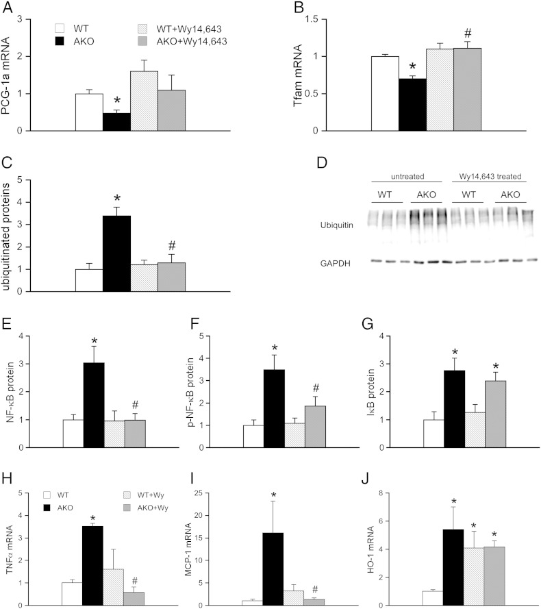
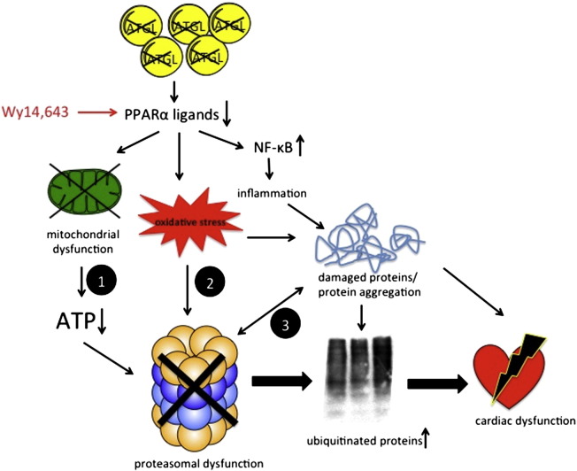
Systemic deletion of the gene encoding for adipose triglyceride lipase (ATGL) in mice leads to severe cardiac dysfunction due to massive accumulation of neutral lipids in cardiomyocytes. Recently, impaired peroxisome proliferator-activated receptor α (PPARα) signaling has been described to substantially contribute to the observed cardiac phenotype. Disturbances of the ubiquitin-proteasome system (UPS) have been implicated in numerous cardiac diseases including cardiomyopathy, ischemic heart disease, and heart failure. The objective of the present study was to investigate the potential role of UPS in cardiac ATGL deficiency. Our results demonstrate prominent accumulation of ubiquitinated proteins in hearts of ATGL-deficient mice, an effect that was abolished upon cardiomyocyte-directed overexpression of ATGL. In parallel, cardiac protein expression of the ubiquitin-activating enzyme E1a, which catalyzes the first step of the ubiquitination cascade, was significantly upregulated in ATGL-deficient hearts. Dysfunction of the UPS was accompanied by activation of NF-κB signaling. Moreover, the endoplasmic reticulum (ER)-resident chaperon protein disulfide isomerase was significantly upregulated in ATGL-deficient hearts. Chronic treatment of ATGL-deficient mice with the PPARα agonist Wy14,643 improved proteasomal function, prevented NF-κB activation and decreased oxidative stress. In summary, our data point to a hitherto unrecognized link between proteasomal function, PPARα signaling and cardiovascular disease.

Keywords: Adipose triglyceride lipase; Cardiac dysfunction; Inflammation; NF-κB; Peroxisome proliferator-activated receptor α.


Figures

Fig. 1
Fig. 1
UPS and cardiac ATGL deficiency. (A) Representative Western blots of ATGL protein expression in cardiac homogenates of WT, AKO, WT/cTg, and AKO/cTg mice. Two different exposure times were chosen to better illustrate supraphysiological ATGL levels in transgene animals. (B) Representative Western blots showing total levels of ubiquitinated proteins as well as UBE1a protein expression in WT, AKO, WT/cTg, and AKO/cTg hearts. (C) Accumulation of ubiquitinated proteins was quantified in cardiac homogenates of WT (open bars), AKO (solid bars), WT/cTg (striped bars), and AKO/cTg (gray bars) mice. Total protein ubiquitination was significantly increased in ATGL deficiency. Data represent mean values ± S.E.M. of 9 individual experiments. (D) Protein expression of UBE1a was upregulated in AKO hearts. Data were expressed as folds of WT control (WT = 1) and represent mean values ± S.E.M. of 6 individual experiments. (E) Chymotrypsin-like proteasomal activity was measured in cardiac cytosols of WT (open circles) and AKO (solid circles) mice over a range of ATP concentrations (0–4 mM). Chymotrypsin-like activity was inhibited in the presence of the peptide aldehyde MG132 (1 μM). (F) Normalization of proteasomal activity to basal values (basal activity = 1) resulted in a similar activation profile. Data represent mean values ± S.E.M. of 5–6 individual experiments; *p < 0.05 vs WT. (G) Protein expression of the 19S regulatory subunit was not affected by ATGL deficiency. (H) Cardiac homogenates of AKO mice showed decreased expression of mitochondrial marker proteins citrate synthetase and prohibitin. Data were expressed as folds of WT control (WT = 1) and represent mean values ± S.E.M. of 6 individual experiments; *p < 0.05 vs WT.
Fig. 2
Fig. 2
Activation of NF-κB signaling in AKO hearts. Protein expression was measured in cardiac homogenates of WT (open bars), AKO (solid bars), WT/cTg (striped bars), and AKO/cTg (gray bars) mice. Protein levels of (A) IKKα but not of (B) IKKβ were significantly increased in ATGL deficiency. Protein expression of (C) NF-κB, (D) p-NF-κB and (E) IκB were raised in AKO hearts. Data were expressed as folds of WT control (WT = 1) and represent mean values ± S.E.M. of 6 individual experiments; *p < 0.05 vs WT; #p < 0.05 vs AKO. (F) Representative Western blots. (G) Cardiac mRNA levels of NF-κB target genes TNFα, MCP-1, IL-6, HO-1, and GTPCH-1 determined by qPCR were upregulated in ATGL deficiency. Data represent mean values ± S.E.M. of 5–10 individual experiments. *p < 0.05 vs WT.
Fig. 3
Fig. 3
Feeding of WT and AKO mice with the PPARα agonist Wy14,643. Protein and mRNA expression was measured in cardiac homogenates prepared from Wy14,643-treated WT (striped bars) and AKO (gray bars) mice and compared to that of non-treated WT (open bars) and AKO (solid bars) controls. Improvement of PPARα signaling by Wy14,643 treatment was confirmed by reversal of (A) PGC-1α and (B) Tfam mRNA expression. Feeding of AKO mice with Wy14,643 reduced cardiac expression of (C, D) ubiquitinated proteins to WT levels. Protein expression of (E) NF-κB and (F) p-NF-κB but not (G) IκB was reversed in Wy14,643-treated AKO mice. Cardiac mRNA levels of the NF-κB target genes (H) TNFα and (I) MCP-1 were reduced to WT levels while increased expression of (J) HO-1 persisted after Wy14,643 treatment. Data represent mean values ± S.E.M. of 5–6 individual experiments. *p < 0.05 vs WT; #p < 0.05 vs AKO.
Fig. 4
Fig. 4
Oxidative stress in Wy14,643-treated AKO mice. Cardiac mRNA expression was measured in homogenates of WT (open bars), AKO (solid bars), Wy14,643-treated WT (striped bars), and Wy14,643-treated AKO/cTg (gray bars) mice. (A) NOX2 mRNA was significantly upregulated in AKO animals. This effect was decreased by treatment with the PPARα agonist Wy14,643. (B) Upregulation of NOX4 mRNA in AKO animals persisted upon Wy14,643 supplementation. (C) Protein expression of p47phox was significantly increased in cardiac homogenates of AKO mice and reduced by 25% upon Wy14,643-treatment. (D) Upregulation of cardiac p67phox protein expression was unaffected by Wy14,643. (E, F) Protein expression of SOD-1 was significantly decreased in untreated and Wy14,643-treated AKO hearts, while cardiac levels of catalase were similar in all experimental groups. Data were expressed as folds of WT control (WT = 1) and represent mean values ± S.E.M. of 6 individual experiments; *p < 0.05 vs WT; #p < 0.05 vs AKO. (G) NADPH oxidase activity was measured as lucigenin-derived chemiluminescence. (H) Representative Western blots.
Scheme 1
Scheme 1
Interplay of signaling pathways leading to cardiac dysfunction. ATGL deficiency in cardiomyocytes results in decreased production of lipid ligands for PPARα activation. Consequent mitochondrial dysfunction results in decreased intracellular ATP levels, leading to reduced activation of the 26S proteasome (path 1). Proteasomal dysfunction causes accumulation and aggregation of ubiquitinated proteins. Increased oxidative inflammatory stress may damage critical residues/domains within the catalytic core of the proteasome (path 2). Damaged and/or misfolded proteins impair proteasomal activity by saturation of the 20S core with non-degradable aggregates (path 3). Restored PPARα signaling with Wy14,643 reduces oxidative inflammatory stress, improves mitochondrial function, and consequently increases UPS activity and cardiac performance.

Similar articles

Cited by

References

    1. Finley D. Recognition and processing of ubiquitin–protein conjugates by the proteasome. Annu Rev Biochem. 2009;78:477–513. doi: 10.1146/annurev.biochem.78.081507.101607. - DOI - PMC - PubMed
    1. Predmore J.M., Wang P., Davis F., Bartolone S., Westfall M.V., Dyke D.B. Ubiquitin proteasome dysfunction in human hypertrophic and dilated cardiomyopathies. Circulation. 2010;121:997–1004. doi: 10.161/CIRCULATIONAHA.109.904557. - DOI - PMC - PubMed
    1. Tsukamoto O., Minamino T., Okada K., Shintani Y., Takashima S., Kato H. Depression of proteasome activities during the progression of cardiac dysfunction in pressure-overloaded heart of mice. Biochem Biophys Res Commun. 2006;340:1125–1133. doi: 10.016/j.bbrc.2005.12.120. - DOI - PubMed
    1. Divald A., Powell S.R. Proteasome mediates removal of proteins oxidized during myocardial ischemia. Free Radic Biol Med. 2006;40:156–164. doi: 10.1016/j.freeradbiomed.2005.09.022. - DOI - PubMed
    1. Powell S.R., Wang P., Katzeff H., Shringarpure R., Teoh C., Khaliulin I. Oxidized and ubiquitinated proteins may predict recovery of postischemic cardiac function: essential role of the proteasome. Antioxid Redox Signal. 2005;7:538–546. doi: 10.1089/ars.2005.7.538. - DOI - PubMed

Publication types

MeSH terms