

Neonatal intrahippocampal HIV-1 protein Tat(1-86) injection: neurobehavioral alterations in the absence of increased inflammatory cytokine activation
- PMID: 25285887
- PMCID: PMC4268159
- DOI: 10.1016/j.ijdevneu.2014.09.004
Neonatal intrahippocampal HIV-1 protein Tat(1-86) injection: neurobehavioral alterations in the absence of increased inflammatory cytokine activation
Abstract
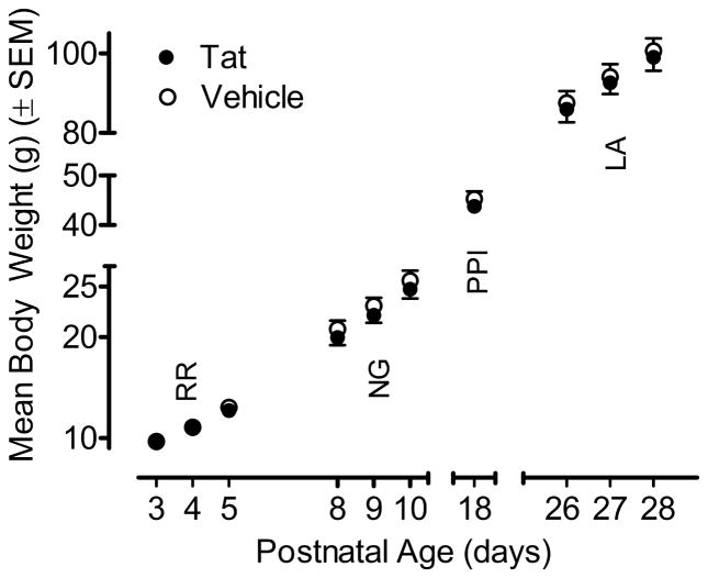
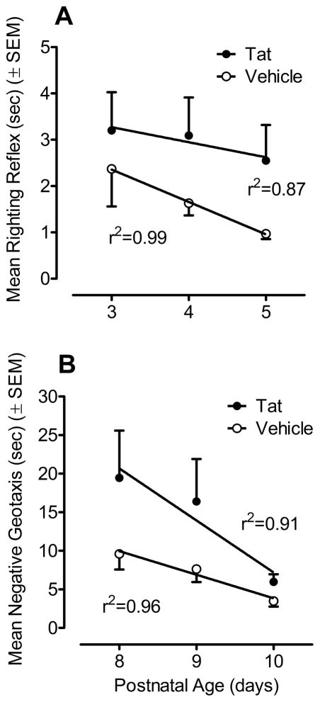
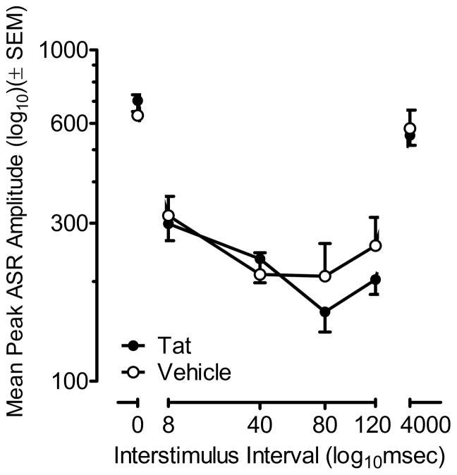
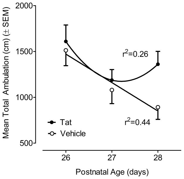
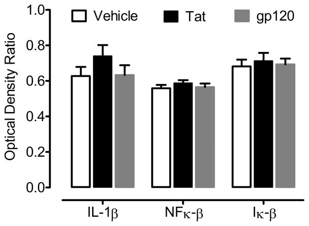
Pediatric AIDS caused by human immunodeficiency virus type 1 (HIV-1) remains one of the leading worldwide causes of childhood morbidity and mortality. HIV-1 proteins, such as Tat and gp120, are believed to play a crucial role in the neurotoxicity of pediatric HIV-1 infection. Detrimental effects on development, behavior, and neuroanatomy follow neonatal exposure to the HIV-1 viral toxins Tat1-72 and gp120. The present study investigated the neurobehavioral effects induced by the HIV-1 neurotoxic protein Tat1-86, which encodes the first and second exons of the Tat protein. In addition, the potential effects of HIV-1 toxic proteins Tat1-86 and gp120 on inflammatory pathways were examined in neonatal brains. Vehicle, 25 μg Tat1-86 or 100 ng gp120 was injected into the hippocampus of male Sprague-Dawley pups on postnatal day 1 (PD1). Tat1-86 induced developmental neurotoxic effects, as witnessed by delays in eye opening, delays in early reflex development and alterations in prepulse inhibition (PPI) and between-session habituation of locomotor activity. Overall, the neurotoxic profile of Tat1-86 appeared more profound in the developing nervous system in vivo relative to that seen with the first exon encoded Tat1-72 (Fitting et al., 2008b), as noted on measures of eye opening, righting reflex, and PPI. Neither the direct PD1 CNS injection of the viral HIV-1 protein variant Tat1-86, nor the HIV-1 envelope protein gp120, at doses sufficient to induce neurotoxicity, necessarily induced significant expression of the inflammatory cytokine IL-1β or inflammatory factors NF-κβ and I-κβ. The findings agree well with clinical observations that indicate delays in developmental milestones of pediatric HIV-1 patients, and suggest that activation of inflammatory pathways is not an obligatory response to viral protein-induced neurotoxicity that is detectable with behavioral assessments. Moreover, the amino acids encoded by the second tat exon may have unique actions on the developing hippocampus.
Keywords: Cytokines; Developmental delay; HIV-1; Neurotoxicity; Tat(1–86); gp120.
Copyright © 2014 ISDN. Published by Elsevier Ltd. All rights reserved.
Figures
Similar articles
-
Neonatal intrahippocampal injection of the HIV-1 proteins gp120 and Tat: differential effects on behavior and the relationship to stereological hippocampal measures.Brain Res. 2008 Sep 26;1232:139-54. doi: 10.1016/j.brainres.2008.07.032. Epub 2008 Jul 17. Brain Res. 2008. PMID: 18674522 Free PMC article.
-
Neonatal intrahippocampal gp120 injection: an examination early in development.Neurotoxicology. 2007 Jan;28(1):101-7. doi: 10.1016/j.neuro.2006.07.014. Epub 2006 Jul 31. Neurotoxicology. 2007. PMID: 16973215 Free PMC article.
-
Dose-dependent neurocognitive deficits following postnatal day 10 HIV-1 viral protein exposure: Relationship to hippocampal anatomy parameters.Int J Dev Neurosci. 2018 Apr;65:66-82. doi: 10.1016/j.ijdevneu.2017.10.009. Epub 2017 Oct 27. Int J Dev Neurosci. 2018. PMID: 29111178 Free PMC article.
-
gp120 as an etiologic agent for NeuroAIDS: neurotoxicity and model systems.Adv Neuroimmunol. 1994;4(3):157-65. doi: 10.1016/s0960-5428(06)80252-4. Adv Neuroimmunol. 1994. PMID: 7874383 Review.
-
Calcium dysregulation and neuronal apoptosis by the HIV-1 proteins Tat and gp120.J Acquir Immune Defic Syndr. 2002 Oct 1;31 Suppl 2:S55-61. doi: 10.1097/00126334-200210012-00005. J Acquir Immune Defic Syndr. 2002. PMID: 12394783 Review.
Cited by
-
Hippocampal Neuronal Loss in Infant Macaques Orally Infected with Virulent Simian Immunodeficiency Virus (SIV).Brain Sci. 2017 Apr 10;7(4):40. doi: 10.3390/brainsci7040040. Brain Sci. 2017. PMID: 28394273 Free PMC article.
-
Of mice and monkeys: can animal models be utilized to study neurological consequences of pediatric HIV-1 infection?ACS Chem Neurosci. 2015 Aug 19;6(8):1276-89. doi: 10.1021/acschemneuro.5b00044. Epub 2015 Jun 19. ACS Chem Neurosci. 2015. PMID: 26034832 Free PMC article. Review.
-
Neurocognitive Complications of Pediatric HIV Infections.Curr Top Behav Neurosci. 2021;50:147-174. doi: 10.1007/7854_2019_102. Curr Top Behav Neurosci. 2021. PMID: 31522375
-
Long-term HIV-1 Tat Expression in the Brain Led to Neurobehavioral, Pathological, and Epigenetic Changes Reminiscent of Accelerated Aging.Aging Dis. 2020 Feb 1;11(1):93-107. doi: 10.14336/AD.2019.0323. eCollection 2020 Feb. Aging Dis. 2020. PMID: 32010484 Free PMC article.
-
Activation of α7 nicotinic acetylcholine receptor ameliorates HIV-associated neurology and neuropathology.Brain. 2021 Dec 16;144(11):3355-3370. doi: 10.1093/brain/awab251. Brain. 2021. PMID: 34196664 Free PMC article.
References
-
- Aksenov MY, Aksenova MV, Nath A, Ray PD, Mactutus CF, Booze RM. Cocaine-mediated enhancement of Tat toxicity in rat hippocampal cell cultures: the role of oxidative stress and D1 dopamine receptor. Neurotoxicology. 2006;27:217–228. - PubMed
-
- Aksenov MY, Hasselrot U, Bansal AK, Wu G, Nath A, Anderson C, Mactutus CF, Booze RM. Oxidative damage induced by the injection of HIV-1 Tat protein in the rat striatum. Neurosci Lett. 2001;305:5–8. - PubMed
-
- Aksenov MY, Hasselrot U, Wu G, Nath A, Anderson C, Mactutus CF, Booze RM. Temporal relationships between HIV-1 Tat-induced neuronal degeneration, OX-42 immunoreactivity, reactive astrocytosis, and protein oxidation in the rat striatum. Brain Res. 2003;987:1–9. - PubMed
-
- Aksenova MV, Aksenov MY, Mactutus CF, Booze RM. Cell culture models of oxidative stress and injury in the central nervous system. Curr Neurovasc Res. 2005;2:73–89. - PubMed
Publication types
MeSH terms
Substances
Grants and funding
LinkOut - more resources
Full Text Sources
Other Literature Sources
Medical
