. 2015 Apr;193(4):1353-59.
doi: 10.1016/j.juro.2014.09.108. Epub 2014 Oct 5.

Canalization of the urethral plate precedes fusion of the urethral folds during male penile urethral development: the double zipper hypothesis

Affiliations

Canalization of the urethral plate precedes fusion of the urethral folds during male penile urethral development: the double zipper hypothesis

Yi Li et al. J Urol. 2015 Apr.

Abstract

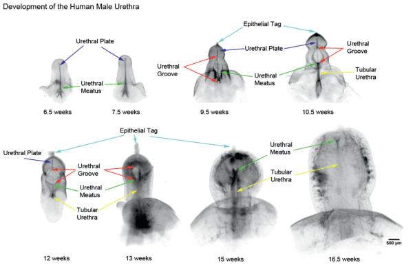
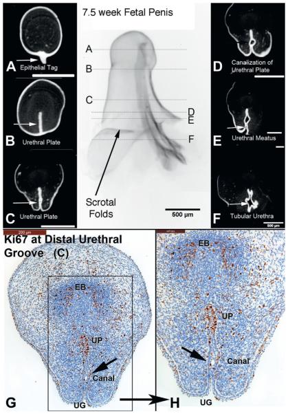
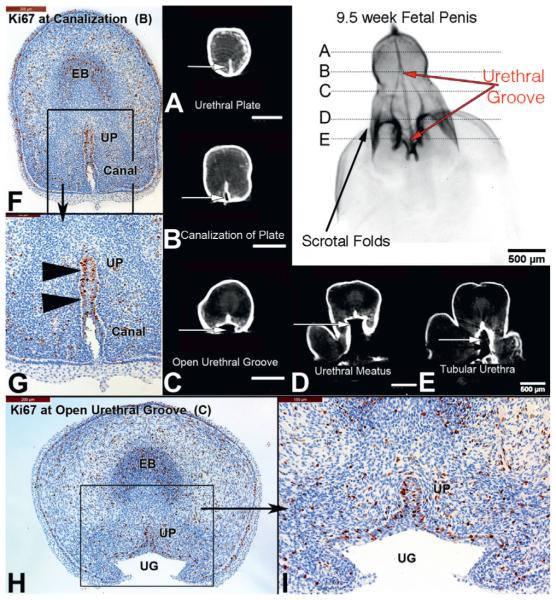
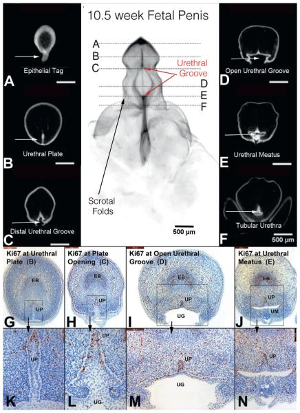
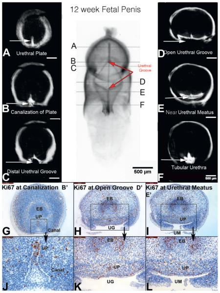
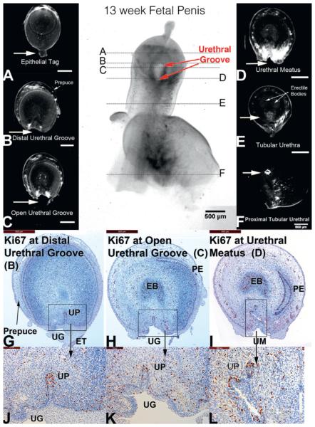
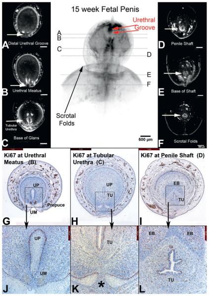
Purpose: We describe the "double zipper" mechanism of human male urethral formation, where the distal zipper opens the urethral groove through canalization of the urethral plate, and a second closing zipper follows behind and closes the urethral groove to form the tubular urethra.

Materials and methods: Anonymous human fetal genital specimens were acquired and gender was determined by polymerase chain reaction of the Y chromosome. Specimens were processed for optical projection tomography, stained with E-cadherin, Ki67 and caspase 3, and imaged.

Results: Eight developing male fetal specimens from 6.5 to 16.5 weeks of gestation were analyzed by optical projection tomography, and an additional 5 specimens by serial sections. Phallus length ranged from 1.3 to 3.7 mm. The urethral plate canalized into a groove with 2 epithelial edges that subsequently fused. Ki67 staining was localized to the dorsal aspect of the urethral plate. In contrast, caspase 3 staining was not observed. The entire process was completed during a 10-week period.

Conclusions: The human male urethra appears to form by 2 mechanisms, an initial "opening zipper" that facilitates distal canalization of the solid urethral plate to form the urethral groove, which involves a high rate of epithelial proliferation (apoptosis not observed), and a "closing zipper" facilitating fusion of the 2 epithelial surfaces of the urethral groove, and thus extending the penile urethra distally. Improved knowledge of the molecular mechanisms of these processes is critical to understanding mechanisms of abnormal urethral development, such as hypospadias.

Keywords: growth and development; hypospadias; organogenesis; urethra.


Figures

Figure 1
Figure 1
OPT of male urethral development from 6.5 to 16.5 weeks of gestation. Note progression of urethral meatus (green arrows) from scrotal folds at 6.5 weeks to terminal position on glans at 16.5 weeks. Wide open urethral groove (red arrows) is best seen from 9.5 to 13 weeks, with clear progression of proximal to distal fusion of edges of urethral groove to form tubular urethra (yellow arrows). At 13 weeks urethral groove is within glans penis with tubular urethra completely formed within shaft of penis, consistent with endodermal theory of urethral development. No evidence of ectodermal intrusion is evident in any specimen.
Figure 2
Figure 2
A to F, OPT of 6.5-week penis at indifferent stage of development stained with E-cadherin. G to K, histological sections correspond to horizontal lines in gross OPT specimen. G and H, note extensive proliferation (Ki67) along urethral plate (UP), urethral meatus (UM) and areas of urethral plate canalization (Canal), ie “opening zipper.” I and J, high power images of G and H. K, caspase 3 was not expressed, in contrast to Ki67 in I and J, supporting role of cellular proliferation over apoptosis as means of urethral plate/groove development. EB, erectile body.
Figure 3
Figure 3
A to F, OPT of 7.5-week penis at indifferent stage of development stained with E-cadherin. G and H, histological sections correspond to horizontal lines in gross specimen. D, E, G and H, note canalization (Canal) of urethral plate (UP) and cellular proliferation (Ki67) localized to dorsal urethral plate (opening zipper). EB, erectile body. UG, urethral groove.
Figure 4
Figure 4
A to E, OPT of 9.5-week penis in early stages of development. B and F to I, urethral groove (UG) is now well developed comprising length of penile shaft (red arrows), and canalization (Canal) of urethral plate (UP, arrowheads) and cellular proliferation (Ki67) localized to dorsal urethral plate (opening zipper). EB, erectile body.
Figure 5
Figure 5
A to F, OPT of 10.5-week penis in early stages of development with epithelium stained for E-cadherin. Continued proliferation (Ki67) in urethral plate (UP, opening zipper, G and H, high power k and L), open urethral groove (UG, red arrows) and fusion of urethral groove (I and M) to form tubular urethra (J and N). EB, erectile body. UM, urethral meatus.
Figure 6
Figure 6
A to F, OPT of 12-week penis in mid stages of development. Note that proximal urethral groove (UG) has fused (closing zipper) and advanced to distal aspect of shaft of penis, and distal groove (opening zipper) has advanced into glans (red arrows). Canal, canalization. EB, erectile body. UM, urethral meatus. UP, urethral plate.
Figure 7
Figure 7
A to F, OPT of 13-week penis in late stages of development. Urethral groove (UG) is contained within proximal glans and is shorter compared to earlier specimens (red arrows). EB, erectile body. ET, epithelial tag. PE, preputial epithelium. UM, urethral meatus. UP, urethral plate.
Figure 8
Figure 8
A to F, OPT of 15-week penis in late stages of development. Note urethral groove is contained within distal glans and is shorter compared to earlier specimens (red arrows). In A prepuce has fused ventrally (G, H, J, K). Asterisk indicates fusion of preputial folds. EB, erectile body. TU, tubular urethra. UM, urethral meatus. UP, urethral plate.

Comment in

  • Editorial comment.
    Kalfa N. Kalfa N. J Urol. 2015 Apr;193(4):1359-60. doi: 10.1016/j.juro.2014.09.128. Epub 2014 Dec 26. J Urol. 2015. PMID: 25545422 No abstract available.

References

    1. Baskin LS, Ebbers MB. Hypospadias: anatomy, etiology, and technique. J Pediatr Surg. 2006;41:463. - PubMed
    1. Glenister TW. A correlation of the normal and abnormal development of the penile urethra and of the infra-umbilical abdominal wall. Br J Urol. 1958;30:117. - PubMed
    1. Kurzrock EA, Baskin LS, Cunha GR. Ontogeny of the male urethra: theory of endodermal differentiation. Differentiation. 1999;64:115. - PubMed
    1. Baskin LS. Hypospadias and urethral development. J Urol. 2000;163:951. - PubMed
    1. Baskin LS, Erol A, Jegatheesan P, et al. Urethral seam formation and hypospadias. Cell Tissue Res. 2001;305:379. - PubMed

Publication types