. 2014 Oct 6;9(10):e108090.
doi: 10.1371/journal.pone.0108090. eCollection 2014.

Neuroprotection of a novel cyclopeptide C*HSDGIC* from the cyclization of PACAP (1-5) in cellular and rodent models of retinal ganglion cell apoptosis

Affiliations

Neuroprotection of a novel cyclopeptide C*HSDGIC* from the cyclization of PACAP (1-5) in cellular and rodent models of retinal ganglion cell apoptosis

Huanhuan Cheng et al. PLoS One. .

Abstract

Purpose: To investigate the protective effects of a novel cyclopeptide C*HSDGIC* (CHC) from the cyclization of Pituitary adenylate cyclase-activating polypeptide (PACAP) (1-5) in cellular and rodent models of retinal ganglion cell apoptosis.

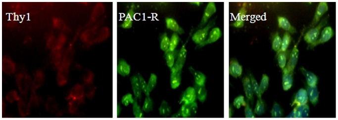
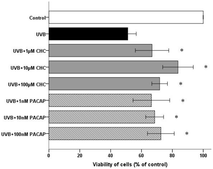
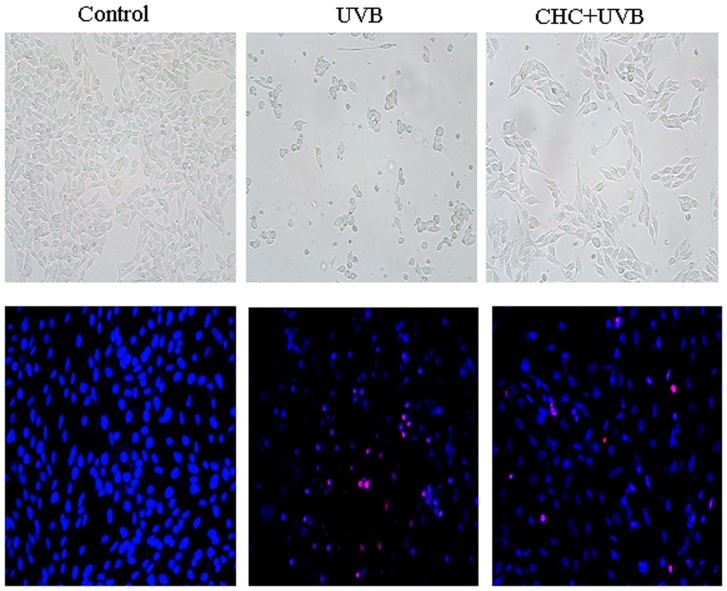
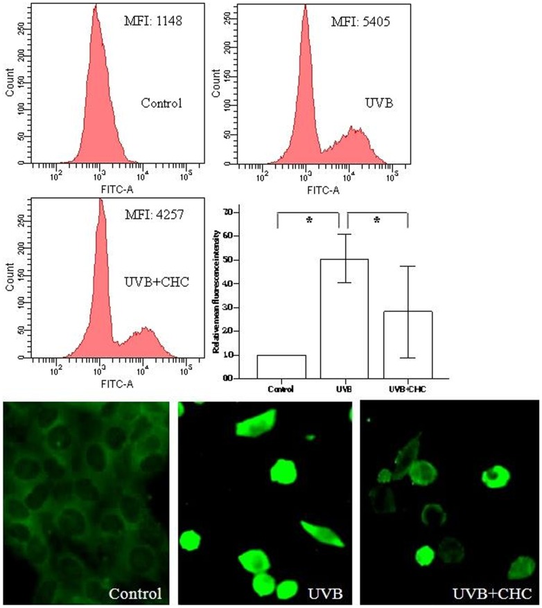
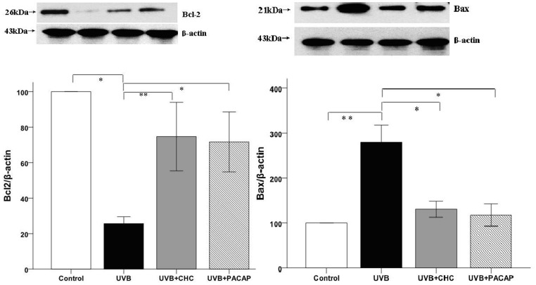
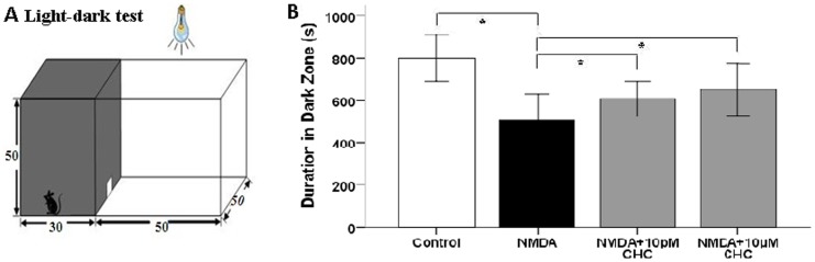
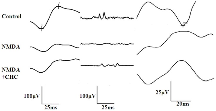
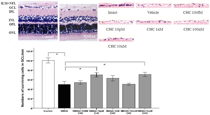
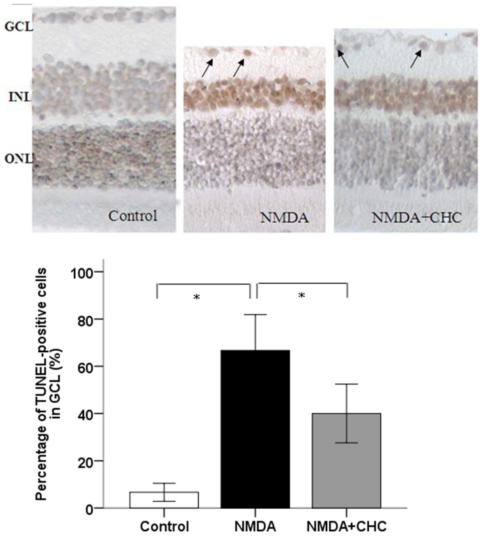
Methodology/principal findings: Double-labeling immunohistochemistry was used to detect the expression of Thy-1 and PACAP receptor type 1 in a retinal ganglion cell line RGC-5. The apoptosis of RGC-5 cells was induced by 0.02 J/cm(2) Ultraviolet B irradiation. MTT assay, flow cytometry, fluorescence microscopy were used to investigate the viability, the level of reactive oxygen species (ROS) and apoptosis of RGC-5 cells respectively. CHC attenuated apoptotic cell death induced by Ultraviolet B irradiation and inhibited the excessive generation of ROS. Moreover, CHC treatment resulted in decreased expression of Bax and concomitant increase of Bcl-2, as was revealed by western-blot analysis. The in vivo apoptosis of retinal ganglion cells was induced by injecting 50 mM N-methyl-D-aspartate (NMDA) (100 nmol in a 2 µL saline solution) intravitreally, and different dosages of CHC were administered. At day 7, rats in CHC+ NMDA-treated groups showed obvious aversion to light when compared to NMDA rats. Electroretinogram recordings revealed a marked decrease in the amplitudes of a-wave, b-wave, and photopic negative response due to NMDA damage. In retina receiving intravitreal NMDA and CHC co-treatment, these values were significantly increased. CHC treatment also resulted in less NMDA-induced cell loss and a decrease in the proportion of dUTP end-labeling-positive cells in ganglion cell line.

Conclusions: C*HSDGIC*, a novel cyclopeptide from PACAP (1-5) attenuates apoptosis in RGC-5 cells and inhibits NMDA-induced retinal neuronal death. The beneficial effects may occur via the mitochondria pathway. PACAP derivatives like CHC may serve as a promising candidate for neuroprotection in glaucoma.


Conflict of interest statement

Competing Interests: The authors have declared that no competing interests exist.

Figures

Figure 1
Figure 1. Immunolocalization studies for Thy-1(red) and PAC1 (green) with double staining.
Evident labeling of the RGC marker Thy-1 was observed in RGC-5 cells. Immunostaining also showed diffuse expression of PAC1 in RGC-5 cells.
Figure 2
Figure 2. Viability of RGC-5 cells in different treatment groups at 24 h post UVB irradiation as measured by MTT assay.
Co-administration of 10 µM CHC conferred the most significant protective effect compared with cells exposed to UVB only. The experiment was carried out three times independently and six replicate wells were set each time. *P<0.05, compared to UVB values.
Figure 3
Figure 3. Representative light micrographs (A–C) and fluorescence micrographs of Hoechst33342 (blue)/PI (red) double staining (D–F) of normal RGC-5 cells, cells subjected to UVB irradiation and cells treated with 10 µM CHC and UVB irradiation.
Normal control cells grew as an interconnected monolayer and exhibited axonal processes (A). Cells after UVB exposure became cuboidal and crenate in shape with the presence of vacuoles (B), which was markedly ameliorated with CHC pretreatment (C). Control cells showed normal nuclear morphology and are negatively stained for PI (D). UVB-irradiated cells including PI-positive cells, showed shrinkage and condensation of their nuclei (E). Treatment with 10 µM CHC reduced both nuclear shrinkage and PI-positive staining (F). Magnification is×200.
Figure 4
Figure 4. Effect of CHC on the ROS level in RGC-5 cells 24 h after UVB exposure as revealed by flowcytometry and fluorescence microscopy.
RGC-5 cells exposed to UVB insult exhibited intense green fluorescent staining. 10 µM CHC clearly blunted the accumulation of ROS in UVB-exposed RGC-5 cells. Data were expressed as means±S.D. (*P<0.05).
Figure 5
Figure 5. Western-blot analysis of RGC-5 cells exposed to UVB irradiation with or without the pretreatment of 10 µM CHC or 100 nM PACAP.
UVB exposure caused a significant decrease in the protein level of Bcl-2 and a concomitant increase of Bax, while CHC significantly decreased the expression of apoptotic marker Bax and increased Bcl-2 expression. The results are expressed as means±S.D. (*P<0.01, **P<0.05).
Figure 6
Figure 6. Enhancement of visual function in NMDA-injected SD rats with CHC co-application.
(A) The light-dark test box consisting of a dark compartment and a larger lit compartment. (B) Time spent in the dark area by four groups of rats. The NMDA-CHC treated rats showed behavioral aversion to light. They spent significantly longer time in the dark chamber than rats injected with NMDA alone (*P<0.01).
Figure 7
Figure 7. Different waves in ERG recordings of different treatment groups.
Representative dark-adapted ERG components (a-wave, b-wave and OPs) and PhNR were recorded in control, NMDA and NMDA+CHC treatment groups. a-wave and b-wave are shown in the first column, while OPs and PhNR were shown in the second and third column respectively.
Figure 8
Figure 8. Representative photomicrograph showing HE staining of cell layers in the retina (100×).
The number of cells per 100 µm in GCL decreased in NMDA-treated retinas but was increased by CHC treatment. The 10 pM and 10 µM CHC -treated groups are significantly different from the control vehicle group. *P<0.01 (ONL outer nuclear layer, OPL outer plexiform layer, INL inner nuclear layer, IPL inner plexiform layer, GCL ganglion cell layer, ILM inner limiting membrane).
Figure 9
Figure 9. Representative photomicrogaphs illustrating the apoptosis of RGCs in response to NMDA (100×).
Deep brown-stained cells indicate TUNEL-positive cells. The proportion of TUNEL-positive cells in the GCL of control SD rats, rats injected with NMDA alone or with NMDA+10 µM CHC were calculated. Data are shown as means±S.E.M. (n = 6). *P<0.05, comparing the NMDA and NMDA+CHC groups.

Similar articles

Cited by

References

    1. Nucci C, Strouthidis NG, Khaw PT (2013) Neuroprotection and other novel therapies for glaucoma. Curr Opin Pharmacol 13: 1–4. - PubMed
    1. Chang EE, Goldberg JL (2012) Glaucoma 2.0: neuroprotection, neuroregeneration, neuroenhancement,. Ophthalmology 119: 979–986. - PMC - PubMed
    1. Yu R, Guo X, Zhong J, Li M, Zeng Z, et al. (2012) The N-Terminal HSDCIF Motif Is Required for Cell Surface Trafficking and Dimerization of Family B G Protein Coupled Receptor PAC1. PLoS ONE 7: e51811. - PMC - PubMed
    1. Vaudry D, Gonzales BJ, Basille M, Yon L, Fournier A, et al. (2000) Pituitary adenylate cyclase activating polypeptide and its receptors: from structure to function. Pharmacol Rev 52: 269–324. - PubMed
    1. Vaudry D, Falluel-Morel A, Bourgault S, Basille M, Burel D, et al. (2009) Pituitary adenylate cyclase activating polypeptide and its receptors: 20 years after thediscovery. Pharmacol Rev 61: 283–357. - PubMed

Publication types

MeSH terms

Substances