. 2014 Oct;70(Pt 10):2640-51.
doi: 10.1107/S1399004714017052. Epub 2014 Sep 27.

Structural analysis of arabinose-5-phosphate isomerase from Bacteroides fragilis and functional implications

Affiliations

Structural analysis of arabinose-5-phosphate isomerase from Bacteroides fragilis and functional implications

Hsiu Ju Chiu et al. Acta Crystallogr D Biol Crystallogr. 2014 Oct.

Abstract

The crystal structure of arabinose-5-phosphate isomerase (API) from Bacteroides fragilis (bfAPI) was determined at 1.7 Å resolution and was found to be a tetramer of a single-domain sugar isomerase (SIS) with an endogenous ligand, CMP-Kdo (cytidine 5'-monophosphate-3-deoxy-D-manno-oct-2-ulosonate), bound at the active site. API catalyzes the reversible isomerization of D-ribulose 5-phosphate to D-arabinose 5-phosphate in the first step of the Kdo biosynthetic pathway. Interestingly, the bound CMP-Kdo is neither the substrate nor the product of the reaction catalyzed by API, but corresponds to the end product in the Kdo biosynthetic pathway and presumably acts as a feedback inhibitor for bfAPI. The active site of each monomer is located in a surface cleft at the tetramer interface between three monomers and consists of His79 and His186 from two different adjacent monomers and a Ser/Thr-rich region, all of which are highly conserved across APIs. Structure and sequence analyses indicate that His79 and His186 may play important catalytic roles in the isomerization reaction. CMP-Kdo mimetics could therefore serve as potent and specific inhibitors of API and provide broad protection against many different bacterial infections.

Keywords: Gram negative; Kdo (3-deoxy-d-manno-oct-2-ulosonate); arabinose 5-phosphate; lipopolysaccharide; structural genomics; sugar isomerase.


Figures
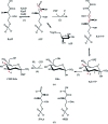
Figure 1
Figure 1
The Kdo biosynthetic pathway, substrate analogues and inhibitors. (a) The Kdo biosynthetic pathway. The enzymes involved are (1) arabinose-5-phosphate isomerase (API), (2) Kdo-8-phosphate (Kdo-8-P) synthase, (3) Kdo-8-P phosphatase and (4) cytidine 5′-monophosphate-Kdo synthetase. API catalyzes the reversible isomerization of d-ribulose 5-phosphate (Ru5P) to d-arabinose 5-phosphate (A5P) in the first step of Kdo biosynthesis. E. coli contains multiple paralogs of API genes including kdsD, kpsF and gutQ. The open-chain form and the ring form of A5P and Kdo-8-P are shown. PEP stands for phosphoenolpyruvate. The C1 and C2 atoms of A5P are marked with red asterisks. The C4 and C5 atoms of CMP-Kdo derived from C1 and C2 of A5P are also marked with red asterisks. (b) 5-Phospho-d-arabinonate (5PAA), a G6P substrate analog commonly used in enzyme mechanistic studies of PGI, is shown on the left. 4-Phospho-2,3-acetyl-d-erythrose hydroxamate (4PEH) was the most potent API inhibitor among the hydroxamate compounds tested in a study of F. tularensis KdsD (Yep et al., 2011 ▶).
Figure 2
Figure 2
Comparison of API sequences. The consensus-aligned sequences of single-domain and multi-domain APIs were calculated using Consensus (http://coot.embl.de/Alignment/consensus.html) and rendered using ESPript (http://espript.ibcp.fr/ESPript/ESPript/). The invariant His79 and His186 are indicated by red triangles. The serine-rich region involved in sugar-phosphate binding is indicated by open green circles. His187 and Tyr190 are conserved among single-domain APIs and are indicated by blue stars. The following abbreviations are used. Single-domain: the consensus sequence at the 90% sequence-identity level of the top 80 single-domain API sequences identified by HMMER. Multi-domain: the consensus sequence at the 90% sequence-identity level of the top 500 multi-domain API sequences identified by HMMER. E. coli_KdsD: KdsD from E. coli K12 substrain MG1655 (ecAPI). CFT073_KdsD, CFT073_KpsF, CFT073_GutQ and CFT073_c3406: KdsD, KpsF, GutQ and c3406 from E. coli CFT073, respectively. lmSPI, putative sugar-phosphate isomerase from Listeria monocytogenes strain 4b F2365.
Figure 3
Figure 3
Crystal structure of bfAPI. (a) Ribbon diagram of the bfAPI tetramer with CMP-Kdo bound at the active site and each monomer rendered in a different color. Bound CMP-Kdo molecules are shown in ball-and-stick representation, with C, O, N and P atoms colored gray, red, blue and orange, respectively. (b) The arrangement of the subunits in an API tetramer in the same orientation as in (a). Subunit A is shown in green ribbon representation and its molecular surface is shown as a gray mesh. The molecular surfaces of subunits B, C and D of bfAPI are colored blue, magenta and yellow, respectively. The tetramer exhibits 222 symmetry, with three orthogonal twofold axes relating one half (dimer) of the tetramer to the other half. These twofold axes pass through the center of the tetramer. One twofold axis runs vertically and the other one runs horizontally along the view shown. The third twofold axis runs perpendicular to the molecule view, going from the outside to the inside of the view. The active site of subunit A is shown and is located at the interface between subunits A, B and C. The CMP-Kdo molecule and the active-site residues His79(C) and His187(B) are shown in ball-and-stick representation. The Ser/Thr-rich loop from subunit A is colored dark blue. (c) Ribbon diagram of a bfAPI monomer color-coded from the N-terminus (blue) to the C-terminus (red). Helices α1–α9 and strands β1–β5 are indicated. The bound CMP-Kdo is shown in a ball-and-stick representation and the Ser/Thr-rich loop is highlighted by a blue oval. (d) The final model of CMP-Kdo fitted in the experimental map, contoured at 1.6 times the r.m.s. of the map (0.75 e Å−3), calculated with SOLOMON solvent-flattened phases from autoSHARP confirm the quality of CMP-Kdo model.
Figure 4
Figure 4
Comparison of API and SPI structures. (a) Superimposition of the bfAPI structure (gray) onto KdsD from E. coli K-12 MG1655 (ecAPI; PDB entry 2xhz; yellow), the putative sugar-phosphate isomerase from L. monocytogenes strain 4b (lmSPI; PDB entry 3fxa; blue) and 3-hexulose-6-phosphate isomerase from M. jannaschii (mjPHI; PDB entry 1jeo; orange). CMP-Kdo in bfAPI is shown in a green ball-and-stick representation. (b) Comparison of the active sites in the bfAPI, ecAPI, lmSPI and mjPHI structures. His79, His186, Tyr190 and CMP-Kdo in bfAPI are shown in green ball-and-stick representation. The dual conformers of His188 in lmSPI subunit D are shown in blue and His176 of mjPHI is shown in an orange ball-and-stick representation. The histidine residues from ecAPI and ImSPI that are equivalent to His79 of bfAPI are shown in yellow and blue ball-and-stick representations, respectively. Residue names are labeled in green, blue and red for bfAPI, lmSPI and mjPHI, respectively.
Figure 5
Figure 5
Comparison of API and PGI structures. (a) Superposition of the bfAPI and rabbit PGI–5PAA (PDB entry 1g98) structures. 1g98 is shown in gray ribbons and the bfAPI monomers superimposed onto the small and large SIS domains of 1g98 are shown as green and yellow ribbons, respectively. CMP-Kdo from the bfAPI structure and 5PAA from the 1g98 structure are shown in ball-and-stick representation. 5PAA was only bound in the small domain of 1g98. (b) Comparison of the bfAPI and the small domains of the PGI structures shows that His79 and the Ser/Thr-rich region are structurally conserved and Ser64 of bfAPI corresponds to a conserved glutamate in PGI. The phosphate group of each of the bound ligand in the PGI structures interacts with the Ser/Thr-rich loop and overlaps with the carboxylate group in Kdo. bfAPI is shown as green ribbons and the PGIs are shown as gray ribbons. The ligands are shown in ball-and-stick representation with N atoms in blue, O atoms in red, P atoms in orange and C atoms of CMP-Kdo and PGIs in cyan and yellow, respectively. The carbon atoms of the active-site residues in bfAPI and PGIs are colored cyan and yellow, respectively. The PDB code, the substrate/product analogs (in parentheses) and the source organisms of the PGI structures used in the superimposition are 2cxp (A5P), mouse; 1u0f (G6P, glucose 6-­phosphate), mouse; 1hox (F6P, fructose 6-phosphate), rabbit; 1g98 (5PAA, 5-phospho-d-arabinonate), rabbit; 1nuh (5PAA), human; 1gzv (5PAA), Sus scrofa; 2o2c (G6P), Trypanosoma brucei; 2q8n, Thermotoga maritima. Note that for clarity only eight of the 16 PGI structures mentioned in §3.5 are shown.
Figure 6
Figure 6
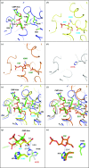
Comparison of bfAPI with mouse PGI–A5P (PDB entry 2cxp), rabbit PGI–F6P (PDB entry 1hox) and mjPHI structures suggests a catalytic role for His79 and His186. The structures of bfAPI, 2cxp, 1hox and mjPHI are shown in ribbon representation and are colored blue, yellow, orange and gray, respectively. CMP-Kdo from bfAPI, A5P from 2cxp, F6P from 1hox and the active-site residues are shown in ball-and-stick representation with N atoms in blue, O atoms in red and P atoms in orange. The C atoms of CMP-Kdo and the active-site residues of bfAPI are in green, while the C atoms of A5P and the active-site residues of 2cxp are in yellow, the C atoms of F6P and the active-site residues of 1hox are in orange and the C atoms of His176 from mjPHI are in gray. Residue names of bfAPI, 2cxp, 1hox and mjPHI are labeled in black, blue, green and red, respectively. (a) The active site of bfAPI. (b) The active site of 2cxp. (c) The active site of 1hox. (d) The active site of the mjPHI structure. (e) Superimposition of the active sites of bfAPI, 2cxp and mjPHI. His79 of bfAPI and His389 of 2cxp are structurally equivalent. The His186 side chain in bfAPI points away from the active site. Ser64 of bfAPI and Glu358 of 2cxp are located in structurally equivalent positions. (f) Superimposition of the active-site structures of bfAPI, 1hox and mjPHI. His79 of bfAPI and His388 of 1hox are structurally equivalent. Ser64 of bfAPI and Glu357 of 1hox are located in structurally equivalent positions. (g) A close-up view of (e). The C1 and C2 atoms of A5P in 2cxp are approximately 3.3 and 2.7 Å away from the N∊2 atom of His176 in mjPHI. If His186 in bfAPI were to adopt a similar side-chain conformation as His176 of mjPHI, it would be placed close to the C1 and C2 atoms of A5P, a position suitable for catalysis. (h) A close-up view of (f). The O5 atom of F6P in 1hox is approximately 2.5 Å away from Nδ1 of His79 of bfAPI. His79 is in a position suitable for catalyzing the ring-opening step. CMP-Kdo was omitted for clarity.

References

    1. Adams, P. D. et al. (2010). Acta Cryst. D66, 213–221. - PubMed
    1. Badger, J. et al. (2005). Proteins, 60, 787–796. - PubMed
    1. Cech, D., Wang, P. F., Holler, T. P. & Woodard, R. W. (2014). J. Bacteriol. 196, 2861–2868. - PMC - PubMed
    1. Cer, R. Z., Mudunuri, U., Stephens, R. & Lebeda, F. J. (2009). Nucleic Acids Res. 37, W441–W445. - PMC - PubMed
    1. Chen, V. B., Arendall, W. B., Headd, J. J., Keedy, D. A., Immormino, R. M., Kapral, G. J., Murray, L. W., Richardson, J. S. & Richardson, D. C. (2010). Acta Cryst. D66, 12–21. - PMC - PubMed

Publication types

MeSH terms

Associated data