. 2014 Oct;70(Pt 10):2740-53.
doi: 10.1107/S1399004714017660. Epub 2014 Sep 27.

Insights into the binding of PARP inhibitors to the catalytic domain of human tankyrase-2

Affiliations

Insights into the binding of PARP inhibitors to the catalytic domain of human tankyrase-2

Wei Qiu et al. Acta Crystallogr D Biol Crystallogr. 2014 Oct.

Abstract

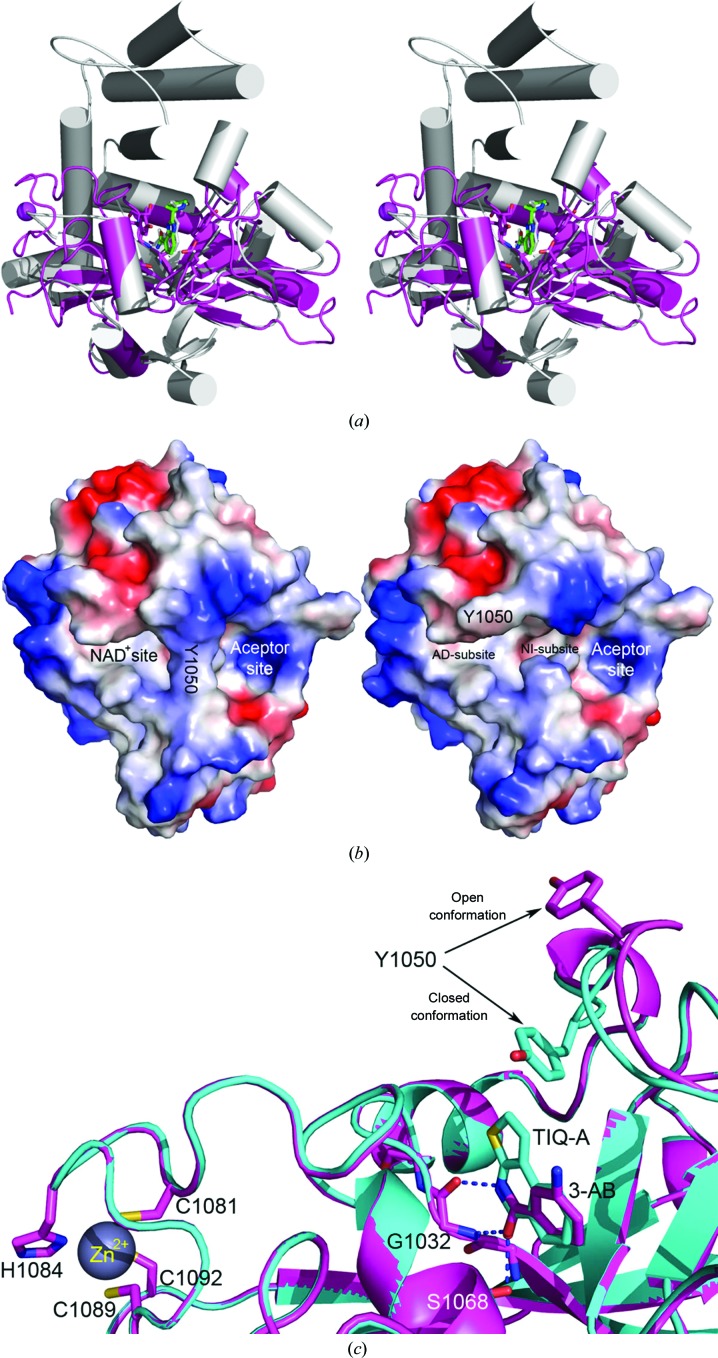
The poly(ADP-ribose) polymerase (PARP) family represents a new class of therapeutic targets with diverse potential disease indications. PARP1 and PARP2 inhibitors have been developed for breast and ovarian tumors manifesting double-stranded DNA-repair defects, whereas tankyrase 1 and 2 (TNKS1 and TNKS2, also known as PARP5a and PARP5b, respectively) inhibitors have been developed for tumors with elevated β-catenin activity. As the clinical relevance of PARP inhibitors continues to be actively explored, there is heightened interest in the design of selective inhibitors based on the detailed structural features of how small-molecule inhibitors bind to each of the PARP family members. Here, the high-resolution crystal structures of the human TNKS2 PARP domain in complex with 16 various PARP inhibitors are reported, including the compounds BSI-201, AZD-2281 and ABT-888, which are currently in Phase 2 or 3 clinical trials. These structures provide insight into the inhibitor-binding modes for the tankyrase PARP domain and valuable information to guide the rational design of future tankyrase-specific inhibitors.

Keywords: TNKS2; cancer; poly(ADP-ribose) polymerase; structural biology; structure-based drug discovery.


Figures

Figure 1
Figure 1
A schematic representation of the TNKS1 and TNKS2 domain architectures and the degrees of sequence identity for the full-length proteins and each domain.
Figure 2
Figure 2
Purified PARP domain of TNKS2 is catalytically active and competent to ribosylate recombinant 3BP2 protein in vitro. An in vitro PARsylation assay was performed using purified PARP domain of TNKS2 and 3BP2 (lanes 4–7) as a substrate or BSA (lane 3) as a control. The PARP inhibitors 3-AB (lane 5), PJ-34 (lane 6) and AZD-2281 (lane 7) were used to inhibit the activity of the PARP domain. Reactions with PARP domain (lane 1) or 3BP2 (lane 2) alone were performed as negative controls. The amount of purified PARP domain and 3BP2/BSA used for reaction was confirmed by Coomassie Blue staining (lower panel).
Figure 3
Figure 3
(a) Stereoview of the crystal structure of the TNKS2–ABT-888 complex (magenta) super­imposed with the structure of PARP2–ABT-888 (gray; PDB entry 3jkd; Karlberg, Hammarström et al., 2010 ▶). (b) The TNKS2 active-site cleft consists of a donor site (NAD+ site) and an acceptor site. The left panel illustrates the closed conformation of the TNKS2 PARP domain. The side chain of Tyr1050 from the D-loop divides the acceptor site and the NAD+ site. The NAD+ site can be further divided into the NI-subsite (where the nicotinamide group is located) and the AD-subsite (which is occupied by the adenosine moiety of NAD+). The right panel represents the opened conformation of the TNKS2 PARP domain, in which the side chain of Tyr1050 swings away from the center of the active site and makes the deeply buried NI-subsite widely accessible. (c) Close-up view of the NI-subsite of TNKS2 in complex with 3-AB (magenta) and TIQ-A (cyan). The side chain of Tyr1050 from the TNKS2–TIQ-A complex is in the closed confirmation compared with the open conformation of the same residue in the TNKS2–3-AB complex. Also, as shown on the far left of the figure, three conserved cysteine residues and one histidine form a short zinc-binding motif involved in the chelation of Zn2+.
Figure 4
Figure 4
(a) The structure of the TNKS2 PARP domain in complex with the first-generation inhibitor 3-AB. 3-AB binds on the bottom of the active site, mimicking the binding mode of nicotinamide. It forms three conserved hydrogen bonds to the backbone of Gly1032 and the side chain of Ser1068. The benzamide ring of 3-AB is in the approximate position to form a π-­stacking interaction with Tyr1071. The 3′ amide of 3-AB forms a connection with the catalytically important residue Glu1138 through a well defined isopropanol molecule (IPA) from the crystallization conditions. The electron density around the inhibitor is a σ-­weighted 2mF oDF c map contoured at 1σ. (b) The B ring of DR-2313 forms three conserved hydrogen bonds to Gly1032 and Ser1068 and a π-stacking interaction with Tyr1071. The A ring also displays hydrophobic interactions with the catalytically important Glu1138 as well as Tyr1060, Phe1061 and Lys1067. The DR-2313 molecule is represented in a stick form covered by spheres, with the S atom colored yellow. (c) NU-1025 forms three hydrogen bonds to Gly1032 and Ser1068 and the π-stacking interaction with Tyr1071 as well as a water-mediated hydrogen bond which links the hydroxyl group of the A ring to Glu1138. (d) 4-HQN forms three hydrogen bonds to Gly1032 and Ser1068 and the π-stacking interaction with Tyr1071. (e) 5-AIQ has similar interactions with TNKS2: three conserved hydrogen bonds to Gly1032 and Ser1068 and a π-stacking interaction with Tyr1071. (f) In addition to the conserved hydrogen bonds and π-stacking interaction, 1,5-IQD makes another water-mediated hydrogen bond from the hydroxyl group of the A ring to Glu1138. (g) TIQ-A forms four hydrogen bonds to TNKS2. The B ring forms three hydrogen bonds to the backbone of Gly1032 and one to the side chain of Ser1068. The tricyclic ring of TIQ-A accounts for a larger planar surface and forms a π-stacking interaction with Tyr1071 compared with the other one-ring or two-ring inhibitors from the same inhibitor class. The side chain of Tyr1050 from the D-loop also swings towards TIQ-A and adopts a closed conformation. (h) INH2BP binds to the NI-subsite differently from the other PARP inhibitors observed in this study. The inhibitor adopts a position in which the iodine moiety points towards the AD-subsite. It does not preserve the three critical hydrogen bonds on the bottom of the NI-subsite observed for 3-AB or TIQ-A. Instead, the hydroxyl group forms only two hydrogen bonds to the main chain of Gly1032 and the side chain of Ser1068.
Figure 5
Figure 5
(a) The tricyclic lactam core of PJ-34 provides multiple contacts within the NI-subsite, with three conserved hydrogen bonds and extended π-sandwich stacking from Tyr1060 and Tyr1071. Unlike the closed conformation observed in the TIQ-A structure (Fig. 4 ▶ g), the tertiary amine extension of PJ-34 pushes the D-loop away from the NAD+ site and the side chain of Tyr1050 adopts an open conformation. (b) The binding features of ABT-888 with TNKS2 compared with the PARP2 catalytic domains. The pyrrolidine ring of ABT-888 is rotated about 10° towards Glu1138 in the TNKS2 structure compared with its position in the PARP2 complex, with a largest shift of 1.7 Å between the two ABT-888 molecules. At the base of the NI-subsite, the carboxamidyl moiety forms three hydrogen bonds to the backbone of Gly1032 and the side chain of Ser1068 in both TNKS2 and PARP2 (the residue numbering is different in PARP2). The N3 atom of the benzimidazole forms a water-mediated hydrogen bond to Glu1138 in the TNKS2 complex (colored magenta), while the N2 atom of the ABT-888 pyrrolidine forms a water-mediated interaction with a glutamate from the N-terminal helix-bundle domain in the PARP2 complex (colored gray). (d) The isoquinolinone base of DPQ contributes to most of the interactions between the inhibitor and TNKS2, with three conserved hydrogen bonds to Gly1032 and Ser1068 as well as π-stacking with Tyr1071 and Tyr1060.
Figure 6
Figure 6
(a) EB-47 occupies the entire NAD-binding pocket in a manner that mimics the binding mode of NAD+. The isoindolinone core interacts with the three conserved hydrogen bonds and a π-stacking effect of Tyr1060 and Tyr1071 can be observed. The adenosine moiety forms four hydrogen bonds to the surrounding protein residues, including an interaction between the hydroxyl group of the ribose and His1031 within the catalytic core. A network of water-mediated hydrogen bonds further enhances the interactions of the compound within the NAD+ donor site. (b) In complex with 3-AB, IWR-1 occupies the AD-subsite without perturbation of the NI-subsite. IWR-1 is stabilized in the AD-subsite primarily through hydrophobic interactions. The adenine ring experiences a π-sandwich stacking interaction between Phe1035 and His1048. The other aspect of the IWR-1 ring structure is its orientation into the hydrophobic pocket surrounded by the side chains of Ile1075, Tyr1071 and Tyr1060. Two additional hydrogen bonds are created to Asp1045. 3-AB is present in the co-crystal near the NI-subsite and contributes to the binding stability of the IWR-1 inhibitor and interacts with Tyr1071 through a π-sandwich stacking interaction, a hydrogen bond to Ser1068 and a water-mediated hydrogen bond to Glu1138. (c) The bicyclic ring of AZD-2281 binds in the NI-subsite by forming the three critical hydrogen bonds and a π-­sandwich stacking interaction. The fluorobenzyl ring in the central position displaces the D-loop by forming a direct hydrogen bond to the backbone of Phe1048. The carbonyl O atom next to the fluorobenzyl ring forms a direct hydrogen bond to the backbone of Tyr1060. Another carbonyl O atom close to the cyclopropyl ring forms one direct hydrogen bond to the backbone of Asp1045 and one water-mediated hydrogen bond to the backbone of Gly1043. The cyclopropyl ring sits in the AD-subsite in between the aromatic rings of Phe1035 and His1048. (d) BSI-201 forms a novel inhibited structure in which two molecules of BSI-201 bind at two distinct sites within the PARP domain of TNKS2. Molecule A of BSI-201 is located in the NI-subsite. The nitro group along with the iodine at the 4-position face towards the center of the protein. The nitro group forms three hydrogen bonds, two with Ser1068 and one with Gly1032, mimicking the function of an amide group. The side chains of Lys1067 and Glu1138 adjust themselves to accommodate the nonpolar interaction with the iodine ion. The amide group on the other side of NI-subsite forms a direct hydrogen bond to the main chain of Gly1032. It also contributes to three water-mediated hydrogen bonds to the main chain of Tyr1071, the main chain of Tyr1060 and the side chain of Ser1033. Another observation is that the side chain of Tyr1050 swings towards BSI-201a to close the NI-subsite in a manner similar to that observed in the TIQ-A complex structure (Figs. 4 ▶ a and 4 ▶ c). Tyr1050 also contributes to the hydrophobic environment for BSI-201a binding. Molecule B of BSI-201 is located in the AD-subsite. The majority of the binding entropy is derived from the π-stacking and nonpolar interactions, with one water-mediated hydrogen bond from the nitro group to the main chain of Asp1045.

Similar articles

Cited by

References

    1. Adams, P. D. et al. (2010). Acta Cryst. D66, 213–221. - PubMed
    1. Afonine, P. V. et al. (2012). Acta Cryst. D68, 352–367. - PMC - PubMed
    1. Ahel, I., Ahel, D., Matsusaka, T., Clark, A. J., Pines, J., Boulton, S. J. & West, S. C. (2008). Nature (London), 451, 81–85. - PubMed
    1. Bauer, P. I., Kirsten, E., Varadi, G., Young, L. J., Hakam, A., Comstock, J. A. & Kun, E. (1995). Biochimie, 77, 374–377. - PubMed
    1. Bonicalzi, M.-E., Haince, J.-F., Droit, A. & Poirier, G. G. (2005). Cell. Mol. Life Sci. 62, 739–750. - PMC - PubMed

Publication types

MeSH terms