. 2014 Dec 1;74(23):7014-23.
doi: 10.1158/0008-5472.CAN-14-1220. Epub 2014 Oct 6.

NGF blockade at early times during bone cancer development attenuates bone destruction and increases limb use

Affiliations

NGF blockade at early times during bone cancer development attenuates bone destruction and increases limb use

Gwen McCaffrey et al. Cancer Res. .

Abstract

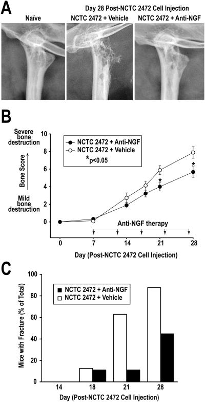
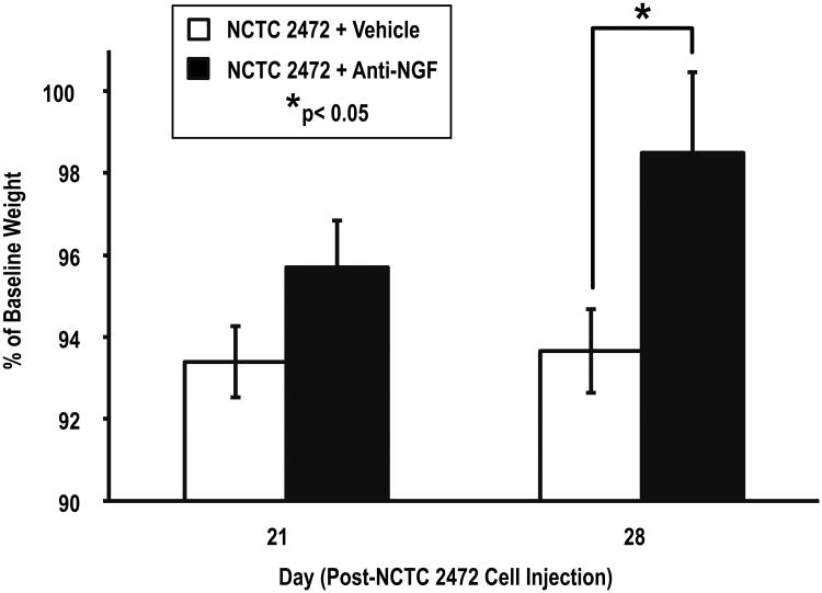
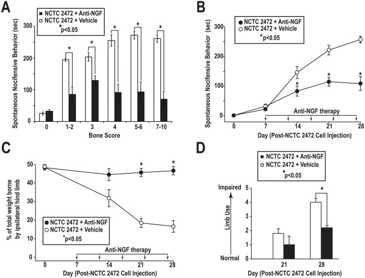
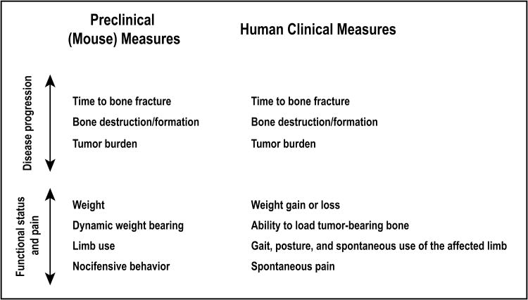
Studies in animals and humans show that blockade of nerve growth factor (NGF) attenuates both malignant and nonmalignant skeletal pain. While reduction of pain is important, a largely unanswered question is what other benefits NGF blockade might confer in patients with bone cancer. Using a mouse graft model of bone sarcoma, we demonstrate that early treatment with an NGF antibody reduced tumor-induced bone destruction, delayed time to bone fracture, and increased the use of the tumor-bearing limb. Consistent with animal studies in osteoarthritis and head and neck cancer, early blockade of NGF reduced weight loss in mice with bone sarcoma. In terms of the extent and time course of pain relief, NGF blockade also reduced pain 40% to 70%, depending on the metric assessed. Importantly, this analgesic effect was maintained even in animals with late-stage disease. Our results suggest that NGF blockade immediately upon detection of tumor metastasis to bone may help preserve the integrity and use, delay the time to tumor-induced bone fracture, and maintain body weight.


Conflict of interest statement

Disclosure of Potential Conflicts of Interest: The authors disclose no potential conflicts of interest.

Figures

Figure 1
Figure 1. Sarcoma-induced bone destruction
High-resolution radiographs of the mouse femur illustrate progressive bone destruction following intramedullary injection of sarcoma cells. To measure the extent of bone destruction, each femur was divided into two equal-length aspects (distal and proximal), and bone destruction scores for each aspect were separately determined. For each femoral aspect, radiographs were scored on a 0-5 scale: 0 (normal), no signs of bone destruction; 1 (mild), small pits (1-3 in number) of bone destruction; 2 (moderate), increased pitted appearance (4-6 in number) and loss of medullary bone; 3 (severe), loss of medullary bone and erosion of cortical bone; 4 (severe + unilateral fracture), full thickness unicortical bone loss; 5 (severe + bilateral fracture), full thickness bicortical bone loss.
Figure 2
Figure 2. Nocifensive behavior over time in bone cancer model
Time-course of spontaneous pain-related behaviors following injection of either NCTC 2472 cells in Hanks balanced salt solution (HBSS) (open circles, n=36) or HBSS (sham) (inverted triangles, n=8) into the mouse femur show that spontaneous nocifensive behavior is temporally separated into “surgical (orthopedic) pain” (Day 1-7) and “bone cancer” pain (Day 7-30). Spontaneous nocifensive behavior exhibited by HBSS-injected (sham) mice show that by Day 7 post-surgery, spontaneous pain-related behaviors approach those exhibited by naïve mice (open squares, n=8). By Day 10 post-cancer-cell injection, time spent in nocifensive behavior by sarcoma-injected mice is easily differentiated from that of sham-injected mice. Error bars represent SEM.
Figure 3
Figure 3. Tumor location (not size) influences cancer pain
To demonstrate that location of the tumor growth (within the bone or within the muscle adjacent but not involving the bone) is the critical factor in the intensity of bone cancer pain, experiments were performed in which murine NCTC 2472 sarcoma-producing cells were injected either directly into the intramedullary space of the mouse femur, or into the muscle directly adjacent to the femur. Representative high-resolution radiographs of a naïve mouse femur (A) a femur from a bone-injected (B), and a muscle-injected (C) mouse at Day 28 post-cancer cell injection revealed the greater tumor volume present in the muscle-injected mouse than in the femur-injected mouse. (D) Tumor volumes dramatically increased following cancer-cell-injection in the muscle-injected mice over time. (E) Despite harboring greater tumor volumes, the time spent in nocifensive behavior by muscle-injected mice (open circles, n=4) was significantly less than that exhibited by femur-injected mice (closed circles, n=6). Error bars represent SEM.
Figure 4
Figure 4. Anti-NGF reduces sarcoma-induced bone destruction
(A) Representative high-resolution radiographs of the proximal aspect of the femur of a naïve mouse, and of vehicle- and anti-NGF-treated mice at Day 28 post-cancer cell injection. Anti-NGF treatment attenuated sarcoma-induced bone destruction. (B) Anti-NGF significantly reduced the extent of bone destruction in therapy-treated mice (closed circles, n=24) compared to vehicle-treated mice (open circles, n=36) at Days 21 and 28 post-cancer cell injection. Error bars represent SEM; *p<0.05, one-way ANOVA. (C) Anti-NGF therapy resulted in an approximate 50% reduction of the number of mice with fractures at Day 28 post-cancer cell injection. Error bars represent SEM.
Figure 5
Figure 5. Anti-NGF therapy attenuates tumor-related weight loss
Mice treated with anti-NGF (closed rectangles, n=24) had significantly less weight loss than vehicle-treated mice (open rectangles, n=36) at Day 28 post-NCTC 2472 cell injection. Error bars represent SEM; *p<0.05, one-way ANOVA.
Figure 6
Figure 6. Anti-NGF attenuates spontaneous and movement-evoked bone cancer pain behaviors
(A) Time spent in spontaneous nocifensive behavior by anti-NGF-treated mice (closed rectangles) with bone scores of 1-2 (minimal bone destruction, early-stage bone cancer disease), 3-4 (moderate bone destruction), and 5-10 (severe to extreme bone destruction with fracture, late-stage bone cancer disease) was significantly reduced as compared to vehicle-treated mice (open rectangles). (B) Anti-NGF-treated mice (closed circles) spent significantly less time in spontaneous nocifensive behavior than vehicle-treated mice (open circles) at Days 14, 21, and 28 post-cancer cell injection. (C) Weight-bearing ability of tumor-bearing hind limbs of anti-NGF-treated mice (closed circles) was preserved throughout the study, whereas the weight-bearing ability of vehicle-treated mice (open circles) was significantly reduced at Days 21 and 28 post-cancer cell injection. (D) Impaired limb use in anti-NGF-treated mice (closed rectangles) was significantly reduced at Day 28 post-cancer cell injection, as compared to vehicle-treated mice (open rectangles). NCTC 2472 + vehicle, n=36; NCTC 2472 + anti-NGF, n=24. Error bars represent SEM; *p<0.05, one-way ANOVA.
Figure 7
Figure 7. Preclinical and clinical measures of bone cancer disease progression, functional status, and cancer-induced bone pain
To facilitate the bench-to-beside translation of preclinical data examining the efficacy of a therapy targeting human bone cancer, it is critical that the pre-clinical animal model incorporate endpoints that are directly translatable to endpoints that can be used in human clinical trials. To that end, we have included in our bone cancer model a variety of preclinical measures of disease progression, functional status, and pain that might be used in human clinical trials involving cancer patients.

References

    1. Jemal A, Bray F, Center MM, Ferlay J, Ward E, Forman D. Global cancer statistics. CA Cancer J Clin. 2011;61:69–90. - PubMed
    1. Ibrahim T, Farolfi A, Mercatali L, Ricci M, Amadori D. Metastatic bone disease in the era of bone-targeted therapy: clinical impact. Tumori. 2013;99:1–9. - PubMed
    1. Cancer.org. American Cancer Society, Inc.; 2014. [updated 2013 Dec 31; cited 2013 Apr 5]. Internet. Available from: http://www.cancer.org/research/cancerfactsstatistics/cancerfactsfigures2....
    1. Farrell C. Bone metastases: assessment, management and treatment options. Br J Nurs. 2013;22:S4, S6, S8–11. - PubMed
    1. von Moos R, Sternberg C, Body JJ, Bokemeyer C. Reducing the burden of bone metastases: current concepts and treatment options. Support Care Cancer. 2013;21:1773–83. - PubMed

Publication types

MeSH terms