Review
. 2014 Dec;105(12):1519-25.
doi: 10.1111/cas.12551. Epub 2014 Nov 5.

Double-strand break repair-adox: Restoration of suppressed double-strand break repair during mitosis induces genomic instability

Affiliations
Review

Double-strand break repair-adox: Restoration of suppressed double-strand break repair during mitosis induces genomic instability

Masahiro Terasawa et al. Cancer Sci. 2014 Dec.

Abstract

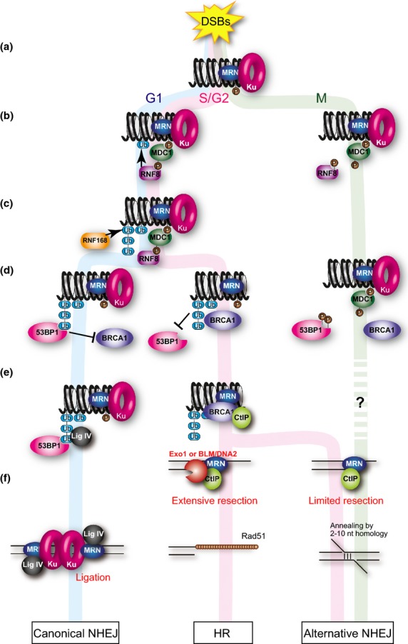
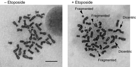
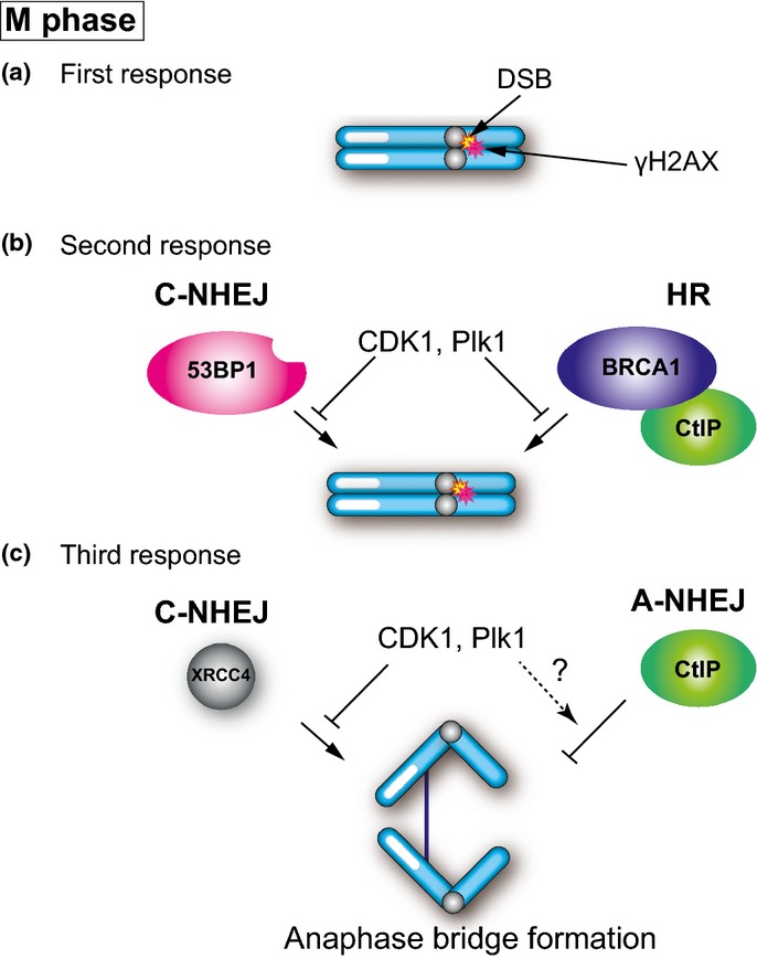
Double-strand breaks (DSBs) are one of the severest types of DNA damage. Unrepaired DSBs easily induce cell death and chromosome aberrations. To maintain genomic stability, cells have checkpoint and DSB repair systems to respond to DNA damage throughout most of the cell cycle. The failure of this process often results in apoptosis or genomic instability, such as aneuploidy, deletion, or translocation. Therefore, DSB repair is essential for maintenance of genomic stability. During mitosis, however, cells seem to suppress the DNA damage response and proceed to the next G1 phase, even if there are unrepaired DSBs. The biological significance of this suppression is not known. In this review, we summarize recent studies of mitotic DSB repair and discuss the mechanisms of suppression of DSB repair during mitosis. DSB repair, which maintains genomic integrity in other phases of the cell cycle, is rather toxic to cells during mitosis, often resulting in chromosome missegregation and aberration. Cells have multiple safeguards to prevent genomic instability during mitosis: inhibition of 53BP1 or BRCA1 localization to DSB sites, which is important to promote non-homologous end joining or homologous recombination, respectively, and also modulation of the non-homologous end joining core complex to inhibit DSB repair. We discuss how DSBs during mitosis are toxic and the multiple safeguard systems that suppress genomic instability.

Keywords: Chromosome segregation; DSB repair; NHEJ; homologous recombination; mitosis.


Figures

Fig. 1
Fig. 1
Role of ubiquitin in the response of double-strand breaks (DSBs) in interphases and mitosis. (a) DSB induces ATM-dependent phosphorylation of histone H2AX. (b) MDC1 recognizes the phosphorylation in both interphase and mitosis. ATM also phosphorylates MDC1 to promote RNF8 recruitment to the DSB sites in interphase. CDK1 phosphorylates RNF8 to inhibit the recruitment during mitosis. (c) RNF8 works with RNF168 to ubiquitinate histone H2A and other molecules to amplify the ubiquitin-mediated DSB signaling. In mitotic cells, the ubiquitination is suppressed. (d) Ubiquitination leads to recruitment of multiple effector proteins such as 53BP1 and BRCA1 in interphases. Both 53BP1 and BRCA1 fail to localize to the DSB sites during mitosis. (e) 53BP1 promotes non-homologous end joining (NHEJ) in G1 phase, whereas BRCA1 promotes homologous recombination (HR) by interacting with CtIP in S/G2 phase. (f) In G1 phase, DSB is ligated by DNA ligase IV, an NHEJ-specific DNA ligase. In S/G2 phase, DSBs are resected by functions of CtIP and the MRN complex to promote Rad51-ssDNA filament formation to execute HR. Alternative NHEJ (A-NHEJ) dependent on CtIP function, which induces limited resection at the DSB site. Cell-cycle regulation of A-NHEJ is not clear.
Fig. 2
Fig. 2
Effect of etoposide-induced double-strand breaks (DSBs) during mitosis on genomic instability of mitotic chromosomes. Representative images of chromosome spreads from etoposide-treated (+Etoposide) and non-treated (−Etoposide) in HCT116 human colon cancer cells arrested in mitosis. Arrows indicate dicentric chromosomes. Arrowheads show fragmented chromosomes. Scale bar = 10 μm.
Fig. 3
Fig. 3
Domain structure of 53BP1 and comparison of CDK1 and PLK1 sites among XRCC4 orthologs. (a) Domain structure and mitosis-specific phosphorylation sites of 53BP1. (b) Domain structure of XRCC4 and conservation of phosphorylation sites among various species. S326 in the C-terminus of human XRCC4 (Hs) is a potential CDK1 phosphorylation site. T222, S256, and S303 are putative PLK1 phosphorylation sites. The gray and black boxes show XLF and DNA ligase IV binding sites, respectively. Phosphorylation of CDK1 or PLK1 sites are shown in mouse (Ms), chicken (Gg), zebrafish (De), and budding yeast (Sc). DNA ligase IV and XLF binding sites in mouse, chicken, and zebrafish were predicted by aligning with the DNA ligase IV and XLF binding sites of human XRCC4 using T-Coffee software.
Fig. 4
Fig. 4
DNA damage response in mitosis. (a) Double-strand break (DSB) induces histone H2AX phosphorylation (γH2AX) by ATM. (b) CDK1 and PLK1 phosphorylate RNF8 and 53BP1 to inhibit 53BP1 and BRCA1 localization of DSB sites. (c) CDK1 and PLK1 phosphorylate XRCC4, a regulatory subunit of the DNA ligase IV complex, to suppress canonical non-homologous end joining (C-NHEJ) activity. CtIP-dependent alternative non-homologous end joining (A-NHEJ) may prevent anaphase bridge formation.

Similar articles

Cited by

References

    1. Chapman JR, Taylor MR, Boulton SJ. Playing the end game: DNA double-strand break repair pathway choice. Mol Cell. 2012;47:497–510. - PubMed
    1. Bogue MA, Wang C, Zhu C, Roth DB. V(D)J recombination in Ku86-deficient mice: distinct effects on coding, signal, and hybrid joint formation. Immunity. 1997;7:37–47. - PubMed
    1. Kabotyanski EB, Gomelsky L, Han JO, Stamato TD, Roth DB. Double-strand break repair in Ku86- and XRCC4-deficient cells. Nucleic Acids Res. 1998;26:5333–42. - PMC - PubMed
    1. Liang F, Romanienko PJ, Weaver DT, Jeggo PA, Jasin M. Chromosomal double-strand break repair in Ku80-deficient cells. Proc Natl Acad Sci USA. 1996;93:8929–33. - PMC - PubMed
    1. McVey M, Lee SE. MMEJ repair of double-strand breaks (director's cut): deleted sequences and alternative endings. Trends Genet. 2008;24:529–38. - PMC - PubMed

Publication types

Substances