. 2014 Dec;82(12):5203-13.
doi: 10.1128/IAI.02083-14. Epub 2014 Oct 6.

Comparative evaluation of lesion development, tissue damage, and cytokine expression in golden hamsters (Mesocricetus auratus) infected by inocula with different Leishmania (Viannia) braziliensis concentrations

Affiliations

Comparative evaluation of lesion development, tissue damage, and cytokine expression in golden hamsters (Mesocricetus auratus) infected by inocula with different Leishmania (Viannia) braziliensis concentrations

Raquel P Ribeiro-Romão et al. Infect Immun. 2014 Dec.

Abstract

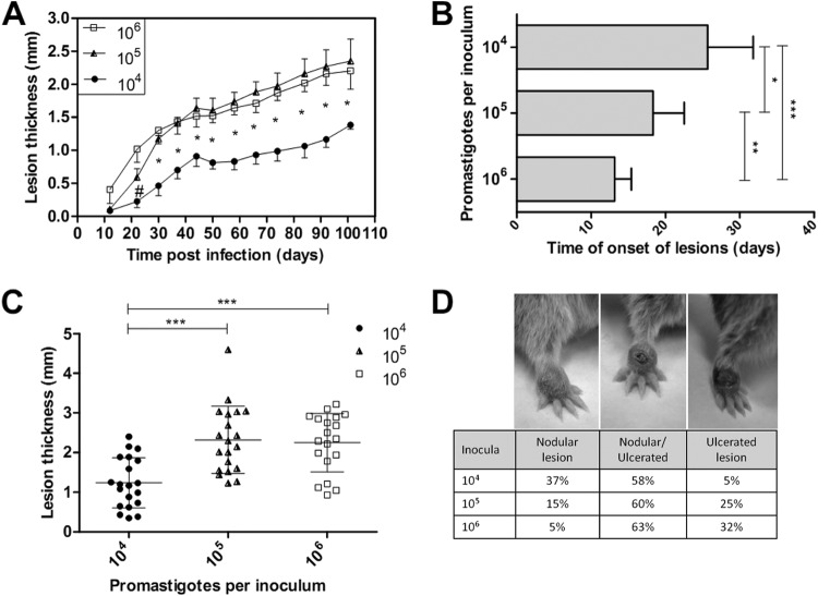
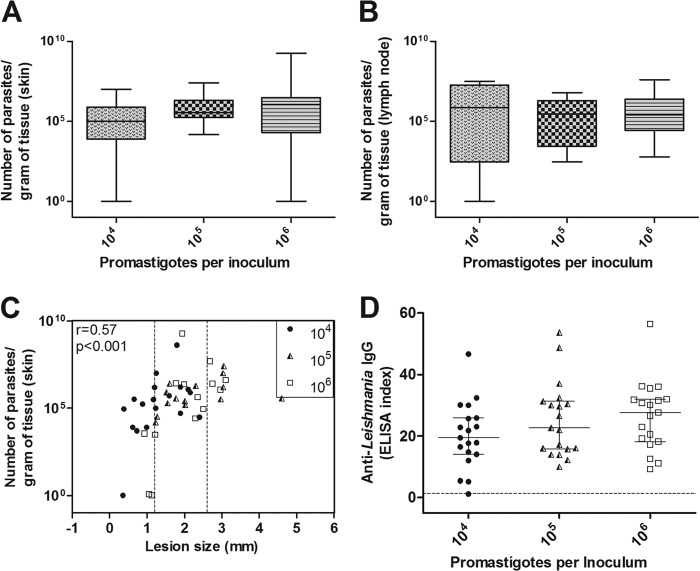
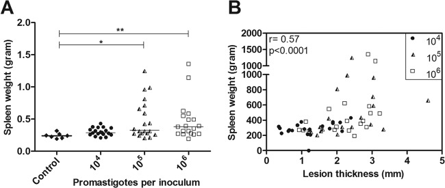
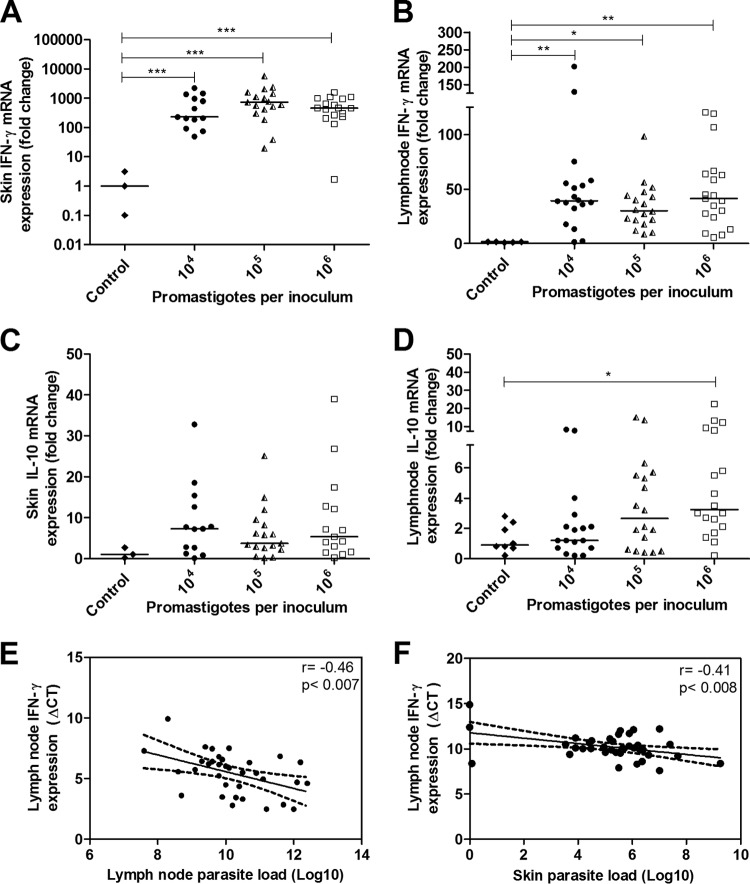
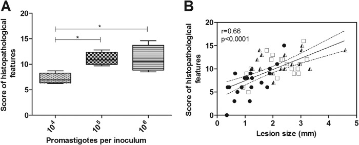
The golden hamster (Mesocricetus auratus) is a susceptible model to Leishmania (Viannia) spp.; however, available studies employ different infection protocols, which account for clinical and pathological presentation differences. Herein, L. (V.) braziliensis preparations were standardized to contain 10(4), 10(5), or 10(6) parasites to determine an optimal inoculum that ensured cutaneous lesions without causing a disseminated infection in hamsters. Lesion development was followed for 105 days by size measurements, and skin, draining lymph node, spleen, and sera were investigated to check parasite load, spleen visceralization, cytokine expression, histopathological changes, and anti-Leishmania IgG levels. The lesion emergence time was inversely proportional to the parasite concentration in the inocula. Animals infected by 10(4) parasites presented nodular lesions, while those infected with 10(6) parasites often exhibited ulcerated lesions. The differences in the final lesion sizes were observed between 10(4) and 10(5) inocula or 10(4) and 10(6) inocula. High IFNG expression, anti-Leishmania IgG levels, and parasite load occurred independently of the inoculum used. A mild inflammatory skin involvement was observed in animals infected with 10(4) parasites, while extensive tissue damage and parasite spleen visceralization occurred with 10(5) and 10(6) parasites. These results indicate that inocula with different concentrations of parasites generate differences in the time of lesion emergence, clinical presentation, and systemic commitment, despite high and similar IFNG expression and parasite load. This suggests that a modulation in the immune response to different parasite numbers occurs in an early phase of the infection, which could dictate the establishment and magnitude of the chronic phase of the disease.


Figures

FIG 1
FIG 1
Evaluation of the golden hamster skin lesions that were infected with Leishmania (Viannia) braziliensis at three different inoculum concentrations (104, 105, and 106 parasites). The lesion sizes were measured by evaluating the dorsal-ventral thickness differences between the infected and uninfected paws. These data are expressed in millimeters. (A) Skin lesion development kinetics through the 105 days following the infections. The graph represents four independent experiments (median and standard error; n = 59 animals). (B) The onset of clinical skin lesions for each inoculum concentration, given in days postinfection (mean ± SD); (C) final skin lesion measurements 105 days after infection (mean ± SD; n = 59 animals; P < 0.0001); (D) final lesion clinical aspect images 105 days after infection. Left, a nodular lesion; middle, a nodular and an ulcerated lesion; right, an ulcerated lesion. #, a significant difference was observed only between the 104 and 106 parasite-infected groups (P < 0.05). *, P < 0.05; **, P < 0.01; ***, P < 0.001.
FIG 2
FIG 2
The parasite load and anti-Leishmania immunoglobulin levels in the golden hamsters that were infected with different Leishmania (Viannia) braziliensis concentrations (104, 105, and 106 parasites) after 105 days of infection. The skin parasite (A) and lymph node parasite (B) loads are shown. (C) The correlation between skin parasite loads and lesion sizes (the medians and interquartile ranges are shown). The dotted lines represent intersection regions in which the lesions that were generated by the three different inocula measured between 1.2 and 2.6 mm (58.7% of the total animals). (D) Anti-Leishmania IgG serum levels. The dotted line represents the cutoff value. Each point represents one animal, and the error bars represent the medians and interquartile ranges (r = correlation coefficient; p = significance level).
FIG 3
FIG 3
The spleen weights in grams from the golden hamsters that were infected with Leishmania (Viannia) braziliensis at three different inoculum concentrations (104, 105, and 106 parasites) after 105 days of infection. (A) The spleen weights in the control and in the three inoculum groups. The horizontal bars represent the medians. *, P < 0.05; **, P < 0.01. (B) Skin lesion sizes positively correlated with spleen weight (Spearman correlation). Each point represents one animal (r = correlation coefficient; p = significance level; *, P < 0.05; **, P < 0.01).
FIG 4
FIG 4
Cytokine mRNA expression as determined by RT-qPCR in the organs of golden hamsters infected with different Leishmania (Viannia) braziliensis parasite concentrations (104, 105, 106 parasites) at 105 days postinfection. The control group represents the uninfected animals. The results are expressed as relative fold changes between the experimental samples and the skin or lymph nodes of a control animal to which the value 1 was arbitrarily assigned. IFN-γ mRNA expression in skin lesions (A) and lymph nodes (B) and IL-10 mRNA expression in skin lesions (C) and lymph nodes (D) are shown. Pearson's correlations between the parasite load and IFN-γ expression in the lymph nodes (E) and the parasite load in skin and the IFN-γ expression in the lymph node (F) are also displayed. Each point represents one animal, and the error bars represent the medians and interquartile ranges. *, P < 0.05; **, P < 0.01; ***, P < 0.001.
FIG 5
FIG 5
Skin lesion histopathological analyses of golden hamsters infected with different Leishmania (Viannia) braziliensis parasite concentrations (104, 105, 106 parasites) at 105 days postinfection. (A) Histopathological features measured by scores in the animal groups infected with 104, 105, and 106 parasites (medians and interquartile ranges) are shown. (B) A correlation between the skin histopathological feature scores and the lesion sizes of the animals infected with 104, 105, and 106 parasites. Each point represents one animal. The correlation graph shows a fit line with a confidence curve. r = correlation coefficient; p = significance level; *, P < 0.05.
FIG 6
FIG 6
Photomicrography of organs from golden hamsters infected with different Leishmania (Viannia) braziliensis concentrations (104, 105, and 106 parasites) at 105 days postinfection. The following are displayed: a localized dermal granuloma that is representative of a 104 parasite-infected animal (hematoxylin and eosin; ×10 magnification) (A), a dermal spread granulomatous reaction that is representative of a 106 parasite-infected animal (arrow, necrosis; arrowhead, Schaumann bodies) (hematoxylin and eosin; ×20 magnification) (B), macrophage vacuoles with amastigotes (arrow) inside a dermal granuloma of a 106 parasite-infected animal (hematoxylin and eosin; ×100 magnification) (C), Schaumann bodies inside a dermal granuloma (arrows) of a 106 parasite-infected animal (hematoxylin and eosin; ×10 magnification) and Schaumann bodies inside a dermal multinucleated giant cell of a 106 parasite-infected animal (inset, ×40 magnification) (D), granulomas in the spleen parenchyma (arrows) (hematoxylin and eosin; ×10 magnification) that are representative of macroscopic splenic nodules of a 106 parasite-infected animal (inset, arrowhead) (E), and splenic vacuolated macrophages that contain Leishmania, which are representative of a 106 parasite-infected animal (arrow) (hematoxylin and eosin; ×100 magnification) (F).

References

    1. Schriefer A, Schriefer AL, Góes-Neto A, Gumarães LH, Carvalho LP, Almeida RP, Machado PR, Lessa HÁ de Jesus AR, Riley LW, Carvalho EM. 2004. Multiclonal Leishmania braziliensis population structure and its clinical implication in a region of endemicity for American cutaneous leishmaniasis. Infect. Immun. 72:508–514. 10.1128/IAI.72.1.508-514.2004. - DOI - PMC - PubMed
    1. Costa JM, Saldanha AC, Nascimento D, Sampaio G, Carneiro F, Lisboa E, Silva ML, Barral A. 2009. Clinical modalities, diagnosis and therapeutic approach of the tegumentary leishmaniasis in Brazil. GM Bahia 79:70–83.
    1. Cupolillo E, Brahim LR, Toaldo CB, de Oliveira-Neto MP, de Brito ME, Falqueto A, de Farias Naiff M, Grimaldi G., Jr 2003. Genetic polymorphism and molecular epidemiology of Leishmania (Viannia) braziliensis from different hosts and geographic areas in Brasil. J. Clin. Microbiol. 41:3126–3132. 10.1128/JCM.41.7.3126-3132.2003. - DOI - PMC - PubMed
    1. Coutinho S, Pirmez C, Mendonça S, Conceição-Silva F, Dórea R. 1987. Pathogenesis in immunopathology of leishmaniasis. Mem. Inst. Oswaldo Cruz 82:214–228. 10.1590/S0074-02761987000500005. - DOI
    1. Jeronimo SM, Duggal P, Ettinger NA, Nascimento ET, Monteiro GR, Cabral AP, Pontes NN, Lacerda HG, Queiroz PV, Gomes CE, Pearson RD, Blackwell JM, Beaty TH, Wilson ME. 2007. Genetic predisposition to self-curing infection with the protozoan Leishmania chagasi: a genomewide scan. J. Infect. Dis. 196:1261–1269. 10.1086/521682. - DOI - PMC - PubMed

Publication types

MeSH terms

LinkOut - more resources