

In vivo study of cerebral white matter in the dog using diffusion tensor tractography
- PMID: 25288360
- PMCID: PMC4409102
- DOI: 10.1111/vru.12211
In vivo study of cerebral white matter in the dog using diffusion tensor tractography
Abstract
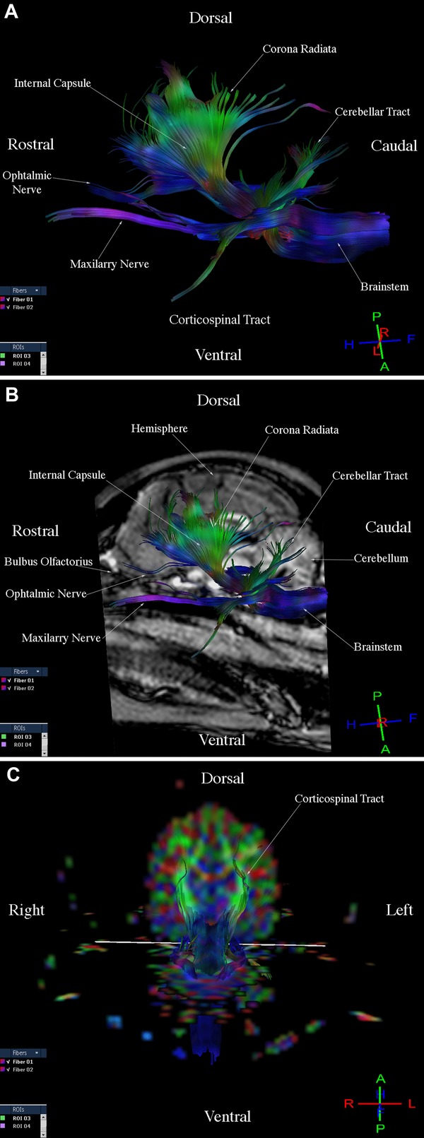
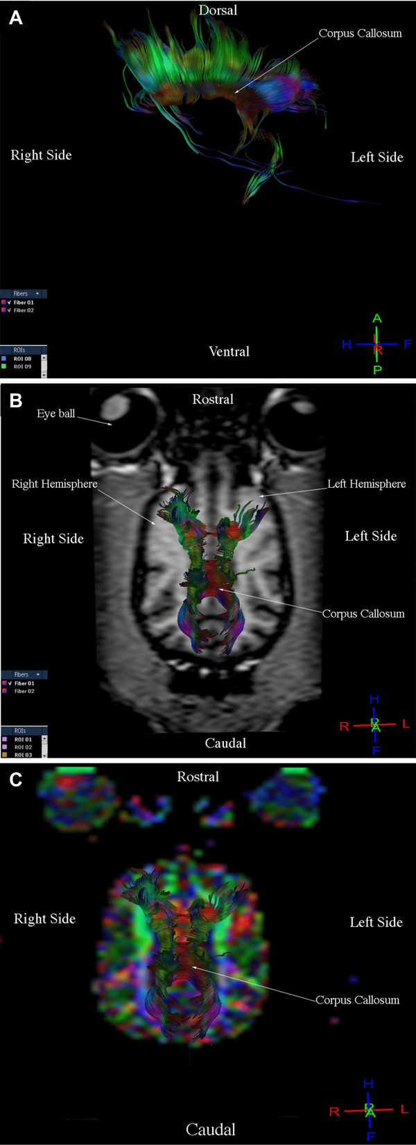
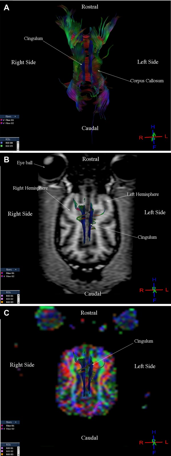
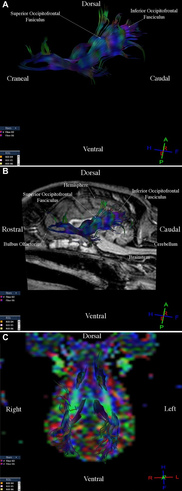
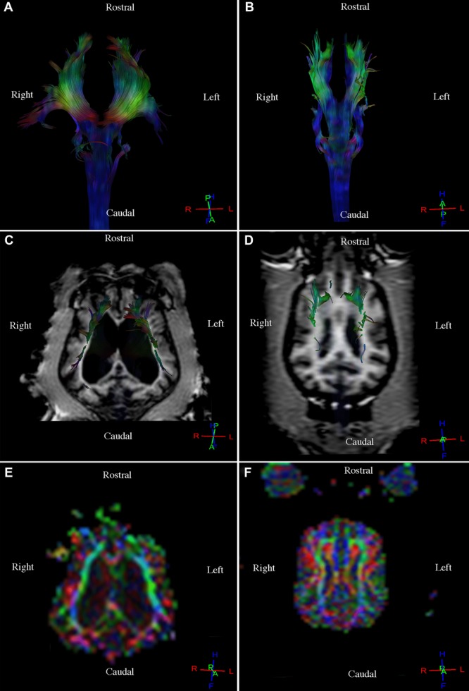
Conventional magnetic resonance imaging (MRI) allows investigators and clinicians to observe the anatomy and injuries of the cerebral white matter (CWM) in dogs. However, dynamic images based on the diffusion tensor (DT) technique are required to assess fiber tract integrity of the CWM. Diffusion tensor tractography (DTT) produces a three-dimensional representation in which data are displayed on a colored map obtained from the anisotropy of water molecules in the CWM tracts. Fractional anisotropy (FA) is a value that measures changes in water diffusion, which can occur if the CWM tracts are displaced, disrupted, or infiltrated. The goal of this study was to determine the feasibility of DTT for in vivo examination of the normal appearance of CWM in dogs through visual and quantitative analysis of the most representative CWM tracts. Nine tractographies were performed on healthy dogs using a 3T MRI scanner. T1- and T2-weighted images and DTI were acquired at different planes. Using DTT, three-dimensional reconstructions were obtained. Fractional ansisotropy and apparent diffusion coefficient (ADC) values of the right and left corticospinal tracts, corpus callosum, cingulum, and right and left fronto-occipital fasciculus were determined. Tract reconstructions were similar in 8/9 healthy dogs. Values for FA and ADC were similar in all the dogs. In one dog, tract reconstructions were inhomogeneous; these were displaced because it had larger lateral ventricles. Findings indicated that DTT is a feasible technique for in vivo study of CWM in dogs and that it complements information from conventional MRI.
Keywords: cerebral white matter; diffusion tensor; dog; in vivo; tractography.
© 2014 The Authors. Veterinary Radiology & Ultrasound published by Wiley Periodicals, Inc. on behalf of American College of Veterinary Radiology.
Figures
References
-
- Martín-Vaquero P, Da-Costa RC, Echandi RL, Tosti CL, Knopp MV, Sammet S. Magnetic resonance imaging of the canine brain at 3 and 7 T. Vet Radiol Ultrasound. 2011;52:25–32. - PubMed
-
- Preti GM, Di Marzio A, Mastropietro A, et al. Boston, Massachusetts, USA: 2011. Tractographic reconstruction protocol optimization in the rat brain in vivo: towards a normal atlas; pp. 8467–8470. 33rd Annual International Conference of the IEEE EMBS. - PubMed
Publication types
MeSH terms
LinkOut - more resources
Full Text Sources
Other Literature Sources
