

Desiccating stress-induced chemokine expression in the epithelium is dependent on upregulation of NKG2D/RAE-1 and release of IFN-γ in experimental dry eye
- PMID: 25288568
- PMCID: PMC4225173
- DOI: 10.4049/jimmunol.1400016
Desiccating stress-induced chemokine expression in the epithelium is dependent on upregulation of NKG2D/RAE-1 and release of IFN-γ in experimental dry eye
Abstract
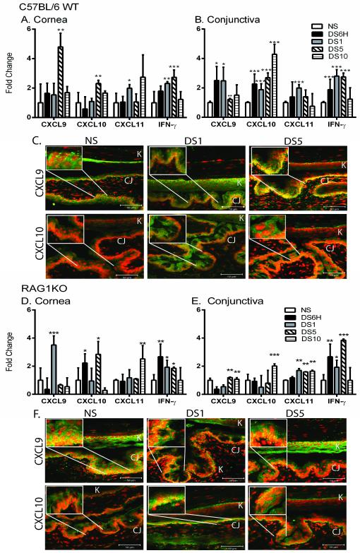
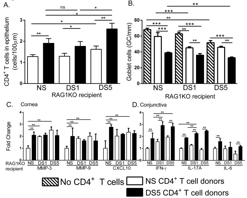
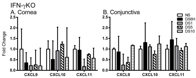
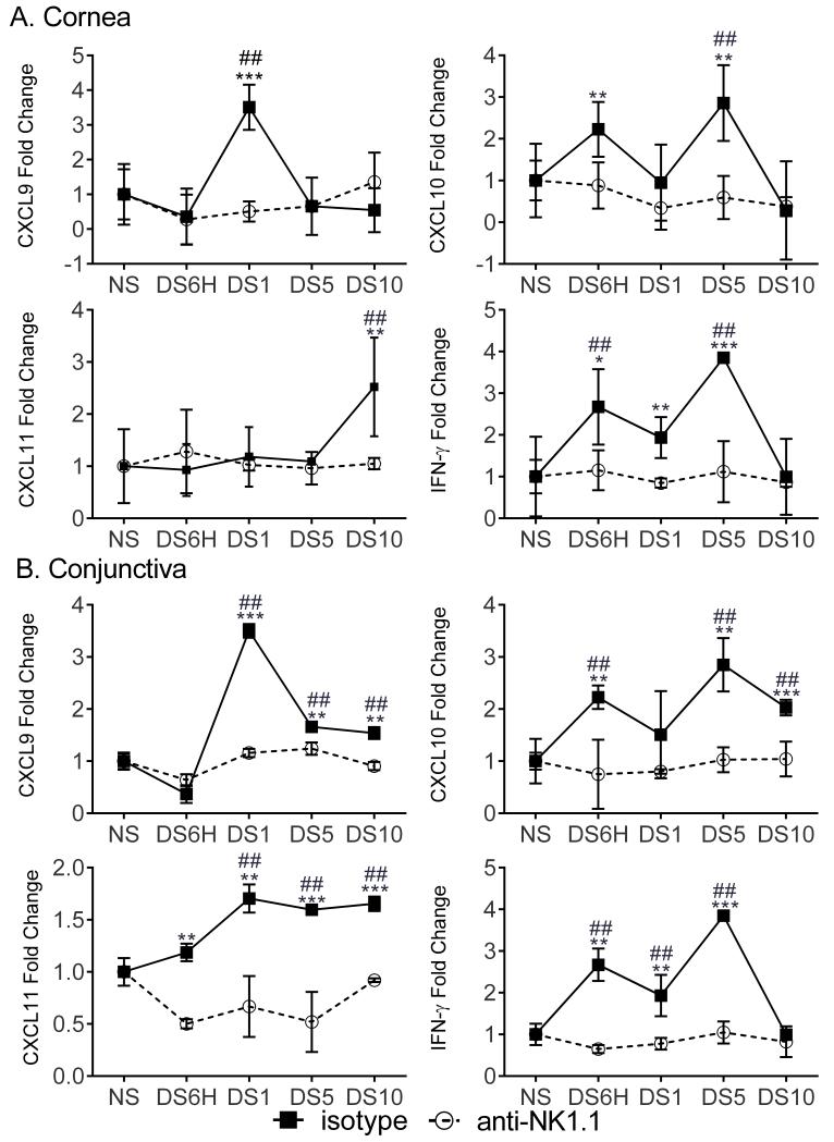
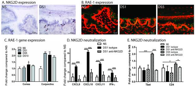
The Th1-associated chemokines CXCL9, CXCL10, and CXCL11 coordinate migration of CXCR3(+) Th1 cells. The objective of this study was to evaluate the role of the innate immune system in stimulating chemokine expression in an experimental model of dry eye and bridge the gap between innate and adaptive immunity. Desiccating stress (DS) induced very early (6 h) expression and production of Th1-associated chemokines in cornea and conjunctiva of C57BL/6 and RAG1 knockout (KO) mice, demonstrating that chemokine expression does not require innate T cells. We then demonstrated that activating the innate immune system prior to adoptive transfer of T cells to RAG1KO mice increased disease severity. Interestingly, lack of induction of chemokines CXCL9, CXCL10, and CXCL11 in IFN-γKO mice provided evidence that their expression requires IFN-γ for induction. Treatment of RAG1KO mice with anti-NK1.1 prevented the increase of CXCL9, CXCL10, and CXCL11 in response to DS, compared with isotype controls. Additionally, DS increased the expression of NKG2D in the conjunctiva. The expression of the NKG2D ligand, retinoic acid early inducible gene 1, also increased at the ocular surface at both the protein and gene levels. Neutralization of NKG2D at the ocular surface decreased the expression of CXCL9, CXCL10, CXCL11, and IFN-γ. In summary, upregulation of CXCL9, CXCL10, and CXCL11 expression in experimental dry eye is T cell-independent, requiring IFN-γ-producing NKG2D(+) NK cells that are activated in response to DS-induced stress signals. This study provides insight into the events that trigger the initial immune response in dry eye pathology.
Copyright © 2014 by The American Association of Immunologists, Inc.
Figures
References
-
- Niederkorn JY, Stern ME, Pflugfelder SC, de Paiva CS, Corrales RM, Gao J, Siemasko K. Desiccating Stress Induces T Cell-Mediated Sjogren’s Syndrome-Like Lacrimal Keratoconjunctivitis. J. Immunol. 2006;176:3950–3957. - PubMed
-
- de Paiva CS, Villarreal AL, Corrales RM, Rahman HT, Chang VY, Farley WJ, Stern ME, Niederkorn JY, Li DQ, Pflugfelder SC. Dry Eye-Induced Conjunctival Epithelial Squamous Metaplasia Is Modulated by Interferon-{gamma} Invest Ophthalmol. Vis. Sci. 2007;48:2553–2560. - PubMed
-
- Zhang X, Chen W, de Paiva CS, Volpe EA, Gandhi NB, Farley WJ, Li DQ, Niederkorn JY, Stern ME, Pflugfelder SC. Desiccating Stress Induces CD4(+) T-Cell-Mediated Sjogren’s Syndrome-Like Corneal Epithelial Apoptosis via Activation of the Extrinsic Apoptotic Pathway by Interferon-gamma. Am J Pathol. 2011;179:1807–1814. - PMC - PubMed
Publication types
MeSH terms
Substances
Grants and funding
- EY018888/EY/NEI NIH HHS/United States
- EY020799/EY/NEI NIH HHS/United States
- S10RR024574/RR/NCRR NIH HHS/United States
- P30CA125123/CA/NCI NIH HHS/United States
- P30 AI036211/AI/NIAID NIH HHS/United States
- P30 EY002520/EY/NEI NIH HHS/United States
- R21 EY018888/EY/NEI NIH HHS/United States
- S10 RR024574/RR/NCRR NIH HHS/United States
- EY018090/EY/NEI NIH HHS/United States
- EY002520/EY/NEI NIH HHS/United States
- EY11915/EY/NEI NIH HHS/United States
- P30AI036211/AI/NIAID NIH HHS/United States
- P30 CA125123/CA/NCI NIH HHS/United States
- P30 EY020799/EY/NEI NIH HHS/United States
- R01 EY018090/EY/NEI NIH HHS/United States
- R01 EY011915/EY/NEI NIH HHS/United States
LinkOut - more resources
Full Text Sources
Other Literature Sources
Research Materials
Miscellaneous
