. 2014 Oct 21;111(42):15214-9.
doi: 10.1073/pnas.1407087111. Epub 2014 Oct 6.

Host cell entry of Middle East respiratory syndrome coronavirus after two-step, furin-mediated activation of the spike protein

Affiliations

Host cell entry of Middle East respiratory syndrome coronavirus after two-step, furin-mediated activation of the spike protein

Jean Kaoru Millet et al. Proc Natl Acad Sci U S A. .

Abstract

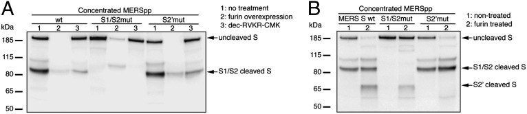
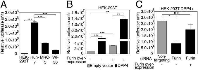
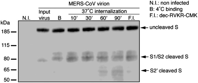
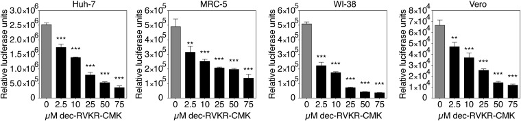
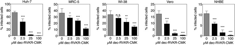
Middle East respiratory syndrome coronavirus (MERS-CoV) is a newly identified betacoronavirus causing high morbidity and mortality in humans. The coronavirus spike (S) protein is the main determinant of viral entry, and although it was previously shown that MERS-CoV S can be activated by various proteases, the details of the mechanisms of proteolytic activation of fusion are still incompletely characterized. Here, we have uncovered distinctive characteristics of MERS-CoV S. We identify, by bioinformatics and peptide cleavage assays, two cleavage sites for furin, a ubiquitously expressed protease, which are located at the S1/S2 interface and at the S2' position of the S protein. We show that although the S1/S2 site is proteolytically processed by furin during protein biosynthesis, the S2' site is cleaved upon viral entry. MERS-CoV pseudovirion infection was shown to be enhanced by elevated levels of furin expression, and entry could be decreased by furin siRNA silencing. Enhanced furin activity appeared to partially override the low pH-dependent nature of MERS-CoV entry. Inhibition of furin activity was shown to decrease MERS-CoV S-mediated entry, as well as infection by the virus. Overall, we show that MERS-CoV has evolved an unusual two-step furin activation for fusion, suggestive of a role during the process of emergence into the human population. The ability of MERS-CoV to use furin in this manner, along with other proteases, may explain the polytropic nature of the virus.

Keywords: Middle East respiratory syndrome coronavirus; furin; proteolytic activation; spike protein; virus entry.


Conflict of interest statement

The authors declare no conflict of interest.

Figures

Fig. 1.
Fig. 1.
Furin proteolytic processing of MERS-CoV S protein. (A) Analysis MERS-CoV S cleavage products during protein biosynthesis. MERS-CoV pseudotyped particles with wt, S1/S2 mutant, or S2′ mutant S were produced in HEK-293T cells that were nontreated (1), overexpressing furin (2), or treated with dec-RVKR-CMK (3). Pseudovirions were harvested and ultracentrifuged. (B) Furin proteolytic processing of MERS-CoV S after virus assembly and egress. MERSpp with wt, S1/S2 mutated, or S2′ mutated S were ultracentrifuged and incubated (2) or not (1) with recombinant human furin. For A and B, samples were analyzed by Western blot, using an anti-S antibody, and arrows denote major S bands observed.
Fig. 2.
Fig. 2.
Role of furin during MERS-CoV S-mediated entry. (A) HEK-293T, Huh-7, MRC-5, and WI-38 cell susceptibility to MERS-CoV S-mediated entry. (B) Effect of enhanced expression of DPP4 receptor and furin on MERSpp entry. HEK-293T cells were transfected with empty vector or DPP4 expression plasmid and cotransfected (+) or not (−) with a furin-encoding plasmid. (C) Effect of furin expression silencing on MERSpp entry. HEK-293T cells were transfected with a DPP4-encoding plasmid and siRNAs that target furin or a nontargeting control siRNA. Cells were cotransfected (+) or not (−) with a furin expression plasmid. For A, B, and C, cells were infected with wt MERSpp, and 72 h postinfection, cells were lysed and luciferase activity was measured. Results are expressed as RLU with error bars representing SD from the mean (n = 3). Data were analyzed using a two-tailed Student t test.
Fig. 3.
Fig. 3.
Furin proteolytic activation of S during internalization of MERS-CoV virions. MERS-CoV virions were bound to the surface of MRC-5 cells at 4 °C for 1 h. Unbound virions were washed and cells were either lysed or incubated for various times (10–90 min) at 37 °C to allow internalization of virions. After each incubation times, cells were lysed. B, binding; N.I., noninfected; F.I., pretreatment with 100 µM dec-RVKR-CMK for 2 h followed by virion binding and internalization for 1 h each. Samples were analyzed by Western blot, using an anti-S antibody. Arrows denote major S bands observed.
Fig. 4.
Fig. 4.
Effect of furin inhibition on MERS-CoV S-mediated entry. Huh-7, MRC-5, WI-38, and Vero cells were pretreated for 2 h with dec-RVKR-CMK at increasing concentrations. The cells were then infected with wt MERSpp. Seventy-two hours postinfection, cells were lysed and luciferase activity was measured. Results are expressed as RLU, with error bars representing SD from the mean (n = 3). Data were analyzed using one-way ANOVA test.
Fig. 5.
Fig. 5.
Effect of furin inhibition on MERS-CoV S infection. Huh-7, MRC-5, WI-38, Vero, and primary NHBE cells were pretreated with increasing concentrations of dec-RVKR-CMK for 2 h. Cells were infected with MERS-CoV at a multiplicity of infection (m.o.i.) of 10 for 8 h (24 h for NHBE). Cells were fixed and immunolabeled for MERS-CoV S and stained for nuclei (DAPI). For each condition, 10× objective fields were randomly acquired and analyzed for total number of cells (DAPI nuclei stain) and S-positive cells (infected cells), with a minimum of 2,400 cells analyzed for each condition. Results are expressed as percentage infected cells, with error bars representing SD from the mean (n = 5). Data were analyzed using one-way ANOVA test.

References

    1. Zaki AM, van Boheemen S, Bestebroer TM, Osterhaus AD, Fouchier RA. Isolation of a novel coronavirus from a man with pneumonia in Saudi Arabia. N Engl J Med. 2012;367(19):1814–1820. - PubMed
    1. van Boheemen S, et al. Genomic characterization of a newly discovered coronavirus associated with acute respiratory distress syndrome in humans. MBio. 2012;3(6):e00473-12. - PMC - PubMed
    1. Alagaili AN, et al. Middle East respiratory syndrome coronavirus infection in dromedary camels in Saudi Arabia. MBio. 2014;5(2):e00884–e14. - PMC - PubMed
    1. Raj VS, et al. Dipeptidyl peptidase 4 is a functional receptor for the emerging human coronavirus-EMC. Nature. 2013;495(7440):251–254. - PMC - PubMed
    1. Chan JF, et al. Differential cell line susceptibility to the emerging novel human betacoronavirus 2c EMC/2012: Implications on disease pathogenesis and clinical manifestation. J Infect Dis. 2013;207(11):1743–52. - PMC - PubMed

Publication types