. 2014 Oct 21;111(42):E4523-31.
doi: 10.1073/pnas.1415279111. Epub 2014 Oct 6.

PGC-1β promotes enterocyte lifespan and tumorigenesis in the intestine

Affiliations

PGC-1β promotes enterocyte lifespan and tumorigenesis in the intestine

Elena Bellafante et al. Proc Natl Acad Sci U S A. .

Abstract

The mucosa of the small intestine is renewed completely every 3-5 d throughout the entire lifetime by small populations of adult stem cells that are believed to reside in the bottom of the crypts and to migrate and differentiate into all the different populations of intestinal cells. When the cells reach the apex of the villi and are fully differentiated, they undergo cell death and are shed into the lumen. Reactive oxygen species (ROS) production is proportional to the electron transfer activity of the mitochondrial respiration chain. ROS homeostasis is maintained to control cell death and is finely tuned by an inducible antioxidant program. Here we show that peroxisome proliferator-activated receptor-γ coactivator-1β (PGC-1β) is highly expressed in the intestinal epithelium and possesses dual activity, stimulating mitochondrial biogenesis and oxygen consumption while inducing antioxidant enzymes. To study the role of PGC-1β gain and loss of function in the gut, we generated both intestinal-specific PGC-1β transgenic and PGC-1β knockout mice. Mice overexpressing PGC-1β present a peculiar intestinal morphology with very long villi resulting from increased enterocyte lifespan and also demonstrate greater tumor susceptibility, with increased tumor number and size when exposed to carcinogens. PGC-1β knockout mice are protected from carcinogenesis. We show that PGC-1β triggers mitochondrial respiration while protecting enterocytes from ROS-driven macromolecule damage and consequent apoptosis in both normal and dysplastic mucosa. Therefore, PGC-1β in the gut acts as an adaptive self-point regulator, capable of providing a balance between enhanced mitochondrial activity and protection from increased ROS production.

Keywords: colon cancer; gene expression; molecular pathology; nuclear receptors.


Conflict of interest statement

The authors declare no conflict of interest.

Figures
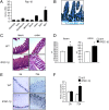
Fig. 1.
Fig. 1.
PGC-1β expression and intestinal morphology. (A) PGC-1β mRNA expression in the gastrointestinal tracts in wild-type mice was measured by real-time qPCR. Results are expressed as mean ± SEM. BAT, brown adipose tissue; WAT, white adipose tissue. (B) Paraffin-embedded ileum specimens from wild-type mice were immunoassayed with PGC-1β antibody to determine expression and localization of the protein. (Magnification: 100×.) (C) Paraffin-embedded ileum and colon specimens from wild-type mice and iPGC-1β mice were stained with H&E and observed by light microscopy. Representative specimens are shown. (Magnification: 100×.) (D) The difference in the dimension of intestinal epithelium was quantified by analyzing the length of crypt–villus axis in the ileum and crypts in the colon (n = 8 mice per group). For each mouse an average of five fields (magnification: 100×) is taken in consideration. The two different groups (n = 10) were compared performed using a Student t test followed by a Mann–Whitney u test. *P < 0.05 was considered significant. (E) Wild-type and iPGC-1β mice were injected i.p. with BrdU (1 mL/100 g body weight) and were killed 2 h or 72 h after injection. Paraffin-embedded ileum specimens from wild-type mice and iPGC-1β mice were immunoassayed with BrdU antibody (Roche Applied Science) to determine the migration of BrdU-positive enterocytes. (Magnification: 100×.) (F) BrdU staining per field was quantified by Image J software and reported as percentage per field. The wild-type and transgenic groups (n = 6) at different time points were compared using a Student t test followed by a Mann–Whitney u test. *P < 0.05 was considered significant.
Fig. 2.
Fig. 2.
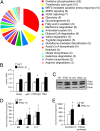
Intestinal PGC-1β induces genes involved in mitochondrial function. (A) The gene-expression profiles of ileum samples from wild-type and iPGC-1β mice were analyzed by microarray analysis. The metabolic pathways differentially expressed in wild-type and iPGC-1β mice were identified using DAVID software available on the DAVID Bioinformatics Resources website (david.abcc.ncifcrf.gov/). The number of genes up-regulated by 1.5 fold in the iPGC-1β mice is indicated for each pathway. (B) Mcad, cytC, ATPβsynt, and mitochondrial Tfam mRNAs were measured in ileum specimens from wild-type and iPGC-1β mice by real-time qPCR. Wild-type and transgenic mice (n = 6) were compared using a Student t test followed by a Mann–Whitney u test. Results are expressed as mean ± SEM; *P < 0.05. (C) Western blot analysis demonstrates increased COXI and porin protein in enterocytes isolated from iPGC-1β mice as compared with enterocytes from wild-type mice. (D) PGC-1β overexpression determines the increase in both the endogenous and COX respiratory capacities of intact enterocytes. A-T, ascorbate/TMPD-dependent oxygen consumption; ER, basal endogenous respiration; UR, DNP-uncoupled respiration. *P < 0.05. (E) Enterocytes isolated from iPGC-1β mice present higher enzymatic activity of both Complex IV and citrate synthase. *P < 0.05.
Fig. 3.
Fig. 3.
Intestinal PGC-1β induces genes involved in antioxidant defense. (A) A mitoSOX assay was performed on enterocytes from wild-type and iPGC-1β mice. The percentage of cells with positive fluorescence cells was normalized with the respective respiratory activities. Results are expressed as mean ± SEM; *P < 0.05. (B) Paraffin-embedded ileum specimens from wild-type mice and iPGC-1β mice were immunoassayed with 8-oxo-dG antibody to determine oxidative stress in the enterocytes and with NITT antibody, a marker of protein damage. The TUNEL assay was performed on paraffin-embedded samples. (Magnification: 200×.) (C) Quantitative analysis of immunostaining by 8-oxo-dG and NITT was performed with Image J software. For the apoptotic TUNEL assay the number of apoptotic cells per crypt–villus unit is indicated. (D) Sod2, Gpx4, Prdx5, Prdx3, and Sirt3 mRNAs were measured in ileum specimens from wild-type mice and iPGC-1β mice by real-time qPCR. Results are expressed as mean ± SEM; *P < 0.05.
Fig. 4.
Fig. 4.
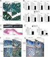
Intestinal PGC-1β promotes chemically and genetically induced intestinal carcinogenesis. (A) Gross morphology of colon samples from wild-type and iPGC-1β mice. (B) Paraffin-embedded ileum and colon specimens from wild-type mice and iPGC-1β mice were stained with H&E and observed by light microscopy. (Magnification: 25×.) (C) Number of tumors (Left) and number of tumors categorized by size (Right) per mouse from colons of wild-type and iPGC-1β mice at the end of the AOM-DSS treatment. The comparison of wild-type and transgenic mice (n = 10) was performed using a Student t test followed by a Mann–Whitney u test. Results are expressed as mean ± SEM; *P < 0.05. (D) Intestinal PGC-1β promotes genetically induced intestinal carcinogenesis. Surviving 7-mo-old iPGC-1β/Apcmin/+ mice presented more tumors in the ileum than FVBN/Apcmin/+ mice. Tumors in in both the ileum and colon were larger in iPGC-1β/Apcmin/+ mice than in FVBN/Apcmin/+ mice. The comparison of FVBN/Apcmin/+ (n = 19) and iPGC-1β/Apcmin/+ mice (n = 12) was performed using a Student t test followed by a Mann–Whitney u test. Results are expressed as mean ± SEM; *P < 0.05.
Fig. 5.
Fig. 5.
Intestinal PGC-1β drives antioxidant enzymes in transformed enterocytes. (A) c-myc levels are similar in tumors from FVBN/Apcmin/+ mice and iPGC-1β/Apcmin/+, but iPGC-1β/Apcmin/+ mice express higher levels of antioxidant enzymes. cytC, Prdx5, Gpx4, Sod2, Sirt3, Txn2, and Prdx3 mRNAs were measured in ileum specimens from wild-type mice and iPGC-1β mice by real-time qPCR. Results are expressed as mean ± SEM; *P < 0.05. (B and C) Paraffin-embedded tumor specimens from FVBN/Apcmin/+ mice and iPGC-1β/Apcmin/+ mice were immunoassayed with PGC-1β and Sod2 (B) or 8-oxo-dG and NITT (C) antibodies. A TUNEL assay was performed on paraffin-embedded samples. (Magnification: 200×.) (D) Quantitative immunostaining analysis for 8-oxo-dG, NITT, and TUNEL was performed with Image J software. *P < 0.05.
Fig. 6.
Fig. 6.
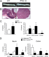
Intestinal PGC-1β ablation decreases antioxidant defense and intestinal carcinogenesis. (A) Paraffin-embedded ileum and colon specimens from PGC-1β fl/? and iPGC-1βKO mice were immunoassayed with PGC-1β antibody to verify its deletion within enterocytes. (Magnification: 100×.) (B) CytC, ATPβsynt, Idh3a, Sirt3, Sod2, Txn2, Prdx3, and Prdx5 mRNAs were measured in colon specimens from PGC-1β fl/? and iPGC-1βKO mice by real-time qPCR. The comparison of wild-type and transgenic mice (n = 6) was performed using a Student t test followed by a Mann–Whitney u test. Results are expressed as mean ± SEM; *P < 0.05. (C) Gross morphology of colon samples from PGC-1β fl/? and iPGC-1βKO mice. (D) Paraffin-embedded ileum and colon specimens from PGC-1β fl/? and iPGC-1βKO mice were stained with H&E and observed by light microscopy. (Magnification: 25×.) (E) Number of tumors (Left) and number of tumors categorized by size (Right) per mouse from colon of PGC-1β fl/? and iPGC-1βKO mice at the end of the AOM-DSS treatment. The comparison of PGC-1β fl/? and iPGC-1βKO mice (n = 10) was performed using a Student t test followed by a Mann–Whitney u test. Results are expressed as mean ± SEM; *P < 0.05. (F and G) Paraffin-embedded ileum specimens from PGC-1β fl/? and iPGC-1βKO mice were immunoassayed with 8-oxo-dG antibody (F) and with NITT antibody (G). Magnification: 100×.
Fig. 7.
Fig. 7.
Transcriptional regulatory network of PGC-1β. (A) Chip-Seq analysis performed with chromatin from enterocytes of wild-type and iPGC-1β mice (Upper) and PGC-1βfl/? and iPGC-1βKO mice (Lower) immunoprecipitated with PGC-1β antibody. The analysis was performed with biological duplicates; results are shown as the sum of binding sites for each group. (B) Venn diagram for TFs whose binding site enrichment is higher in iPGC-1β mice (group 1) or is reduced in iPGC-1βKO mice (group 2) and TFs that were predicted by Ingenuity pathway analysis to be activated (group 3) or inhibited (group 4) in transgenic and knockout intestines, respectively.

References

    1. Ames BN, Shigenaga MK, Gold LS. DNA lesions, inducible DNA repair, and cell division: Three key factors in mutagenesis and carcinogenesis. Environ Health Perspect. 1993;101(Suppl 5):35–44. - PMC - PubMed
    1. D’Errico I, et al. Peroxisome proliferator-activated receptor-gamma coactivator 1-alpha (PGC1alpha) is a metabolic regulator of intestinal epithelial cell fate. Proc Natl Acad Sci USA. 2011;108(16):6603–6608. - PMC - PubMed
    1. St-Pierre J, et al. Suppression of reactive oxygen species and neurodegeneration by the PGC-1 transcriptional coactivators. Cell. 2006;127(2):397–408. - PubMed
    1. Wu Z, et al. Mechanisms controlling mitochondrial biogenesis and respiration through the thermogenic coactivator PGC-1. Cell. 1999;98(1):115–124. - PubMed
    1. Puigserver P, et al. A cold-inducible coactivator of nuclear receptors linked to adaptive thermogenesis. Cell. 1998;92(6):829–839. - PubMed

Publication types

MeSH terms

Substances

Associated data