. 2014 Oct 21;111(42):15155-60.
doi: 10.1073/pnas.1417549111. Epub 2014 Oct 6.

Loss of p53 induces cell proliferation via Ras-independent activation of the Raf/Mek/Erk signaling pathway

Affiliations

Loss of p53 induces cell proliferation via Ras-independent activation of the Raf/Mek/Erk signaling pathway

Matthias Drosten et al. Proc Natl Acad Sci U S A. .

Abstract

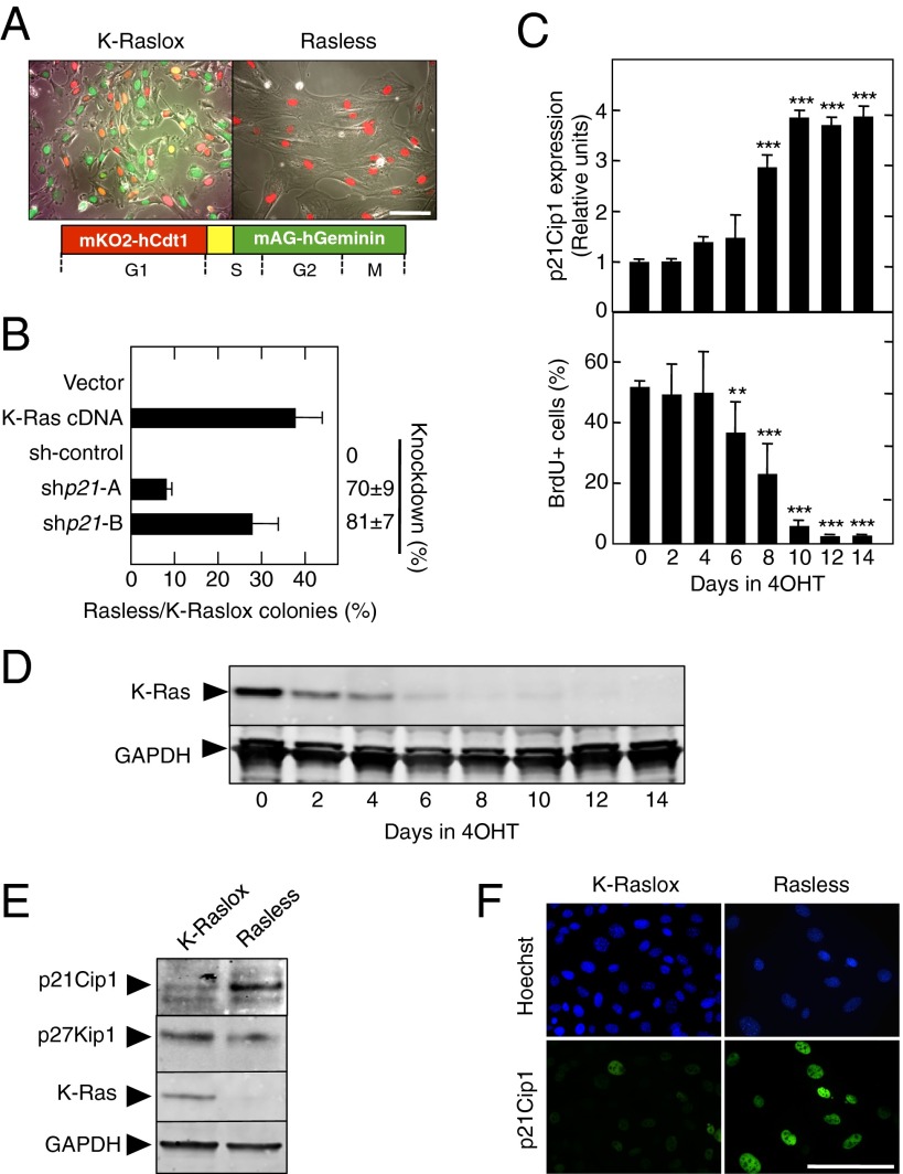
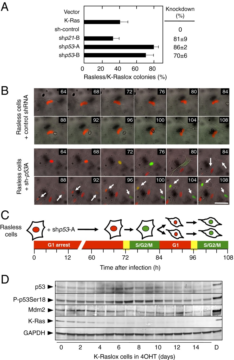
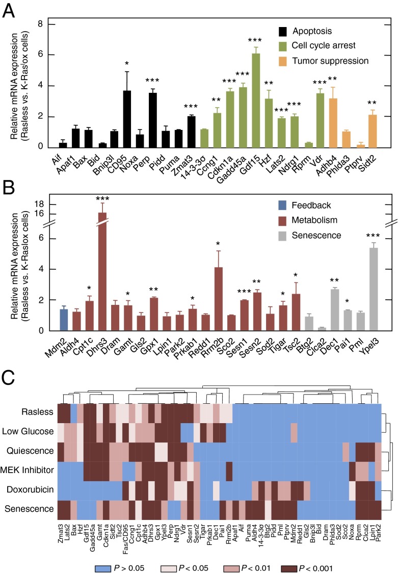
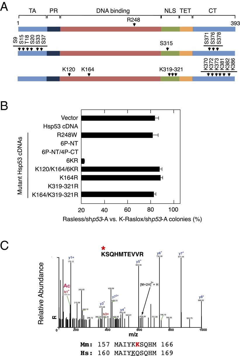
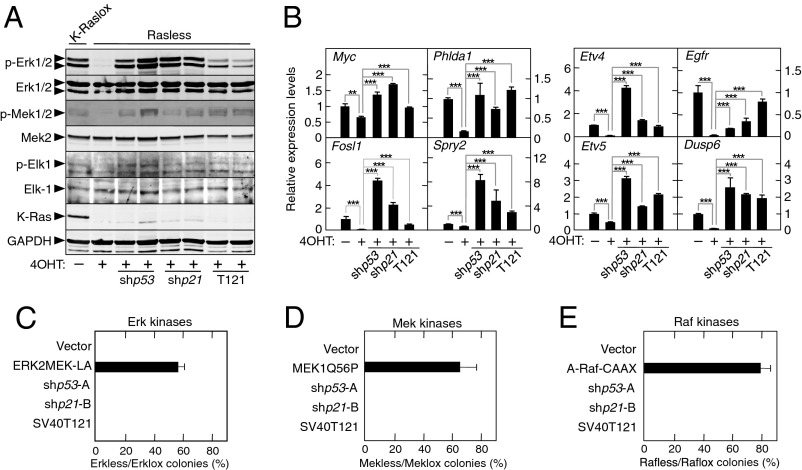
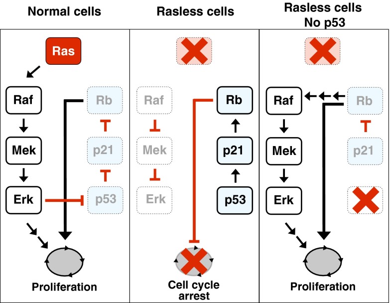
The Ras family of small GTPases constitutes a central node in the transmission of mitogenic stimuli to the cell cycle machinery. The ultimate receptor of these mitogenic signals is the retinoblastoma (Rb) family of pocket proteins, whose inactivation is a required step to license cell proliferation. However, little is known regarding the molecular events that connect Ras signaling with the cell cycle. Here, we provide genetic evidence to illustrate that the p53/p21 Cdk-interacting protein 1 (Cip1)/Rb axis is an essential component of the Ras signaling pathway. Indeed, knockdown of p53, p21Cip1, or Rb restores proliferative properties in cells arrested by ablation of the three Ras loci, H-, N- and K-Ras. Ras signaling selectively inactivates p53-mediated induction of p21Cip1 expression by inhibiting acetylation of specific lysine residues in the p53 DNA binding domain. Proliferation of cells lacking both Ras proteins and p53 can be prevented by reexpression of the human p53 ortholog, provided that it retains an active DNA binding domain and an intact lysine residue at position 164. These results unveil a previously unidentified role for p53 in preventing cell proliferation under unfavorable mitogenic conditions. Moreover, we provide evidence that cells lacking Ras and p53 proteins owe their proliferative properties to the unexpected retroactivation of the Raf/Mek/Erk cascade by a Ras-independent mechanism.


Conflict of interest statement

The authors declare no conflict of interest.

Figures

Fig. 1.
Fig. 1.
p21Cip1 is an essential mediator of cell cycle arrest in the absence of Ras signaling. (A) Cell cycle distribution of K-Raslox (Left) and Rasless (Right) MEFs stably expressing the FUCCI cell cycle indicators monomeric version of Kusabira Orange 2 (mKO2)/human chromatin licensing and DNA replication factor 1 (hCdt1) (red) and monomeric version of Azami Green (mAG)/human geminin (hGeminin) (green). (Lower) Schematic outline of the FUCCI cell cycle indicator system is shown. (B) Colony formation of Rasless and K-Raslox MEFs transfected with the indicated cDNA or shRNAs and expressed as the ratio between the number of colonies observed in cells lacking Ras proteins (Rasless) and those cells expressing K-Ras (K-Raslox). The knockdown efficiency of the shRNAs is indicated on the right side of the graph. Data are represented as mean ± SD. (C, Upper) qRT-PCR showing relative expression levels of p21Cip1 mRNA in K-Raslox MEFs treated for the indicated time with 4OHT. β-Actin expression levels were used for normalization. (C, Lower) Percentages of BrdU+ cells in K-Raslox MEFs treated for the indicated time with 4OHT. Data are represented as mean ± SD. **P < 0.01; ***P < 0.001 (unpaired Student t test). (D) Western blot analysis of K-Ras expression in K-Raslox MEFs treated for the indicated time with 4OHT using anti–Pan-Ras antibodies. GAPDH expression served as a loading control. (E) Western blot analysis of p21Cip1, p27Kip1, and K-Ras expression in K-Raslox MEFs either left untreated (K-Raslox) or treated with 4OHT (Rasless) for 2 wk. GAPDH expression served as a loading control. (F) Immunofluorescence staining of p21Cip1 expression in K-Raslox and Rasless cells. Cells were counterstained with Hoechst 33342 to visualize nuclei. (Scale bars, 100 μm.)
Fig. 2.
Fig. 2.
p53 is required for p21Cip1 expression and cell cycle arrest in the absence of Ras signaling. (A) Colony formation of Rasless and K-Raslox MEFs transfected with the indicated cDNA or shRNAs and expressed as the ratio between the number of colonies observed in cells lacking Ras proteins (Rasless) and those cells expressing K-Ras (K-Raslox). The knockdown efficiency of the shRNAs is indicated on the right side of the graph. Data are represented as mean ± SD. (B) Time-lapse imaging of Rasless cells stably expressing the FUCCI cell cycle indicators mKO2-hCdt1 (red) and mAG-hGeminin (green) infected with lentiviruses expressing a control shRNA (Upper) or shp53-A (Lower). Arrows indicate two daughter cells generated from a Rasless cell that reentered the cell cycle. Time after infection (in hours) is shown in the upper right corner of each photograph. (Scale bar, 50 μm.) (C) Schematic representation of the events shown in B. (D) Western blot analysis of p53, p53 phosphorylated at serine 18 (P-p53Ser18), murine double minute 2 (Mdm2), and K-Ras expression in K-Raslox MEFs left untreated or treated with 4OHT for the indicated times. K-Raslox MEFs treated with 5 μg/mL doxorubicin (D) for 24 h were used as a positive control. GAPDH expression served as a loading control.
Fig. 3.
Fig. 3.
Activation of p53 target genes in Rasless cells. (A and B) qRT-PCR of the indicated p53 target genes in Rasless vs. K-Raslox cells. Genes have been grouped by function, including apoptosis (black bars), cell cycle arrest (green bars), tumor suppression (orange bars), feedback (blue bar), metabolism (red bars), and senescence (gray bars). Data are represented as mean ± SD. *P < 0.05; **P < 0.01; ***P < 0.001 (unpaired Student t test). (C) Heat map of significantly induced genes as determined by qRT-PCR of 50 representative p53 target genes under various stress conditions compared with untreated K-Raslox cells. Stress conditions included loss of Ras gene expression upon incubation of K-Raslox cells with 4OHT for 2 wk (Rasless), exposure to 1 mM glucose for 24 h (Low Glucose), incubation in the presence of 0.1% FBS for 24 h (Quiescence), treatment with 0.4 μM PD0325901 for 24 h (Mek Inhibitor) or with 5 μg/mL doxorubicin for 24 h (Doxorubicin), and replicative senescence of primary K-Raslox MEFs (Senescence). Blue bars, P > 0.05; light brown bars, P < 0.05; medium brown bars, P < 0.01; dark brown bars, P < 0.001 (Student t test).
Fig. 4.
Fig. 4.
Requirement for p53 acetylation for cell cycle arrest in the absence of Ras signaling. (A) Schematic representation of the Hsp53 residues mutated in this study. Residues involved in DNA binding (Top), phosphorylation (Middle), and acetylation (Bottom) events are indicated. Functional domains of p53, including the transactivation domain (TA), the proline-rich region (PR), the DNA binding domain (DNA binding), the nuclear localization signal (NLS), the tetramerization domain (TET), and the C-terminal region (CT), are indicated. (B) Colony formation of Rasless and K-Raslox MEFs expressing the shp53-A shRNA and infected with an empty retrovirus (Vector) or retroviruses encoding the indicated cDNAs and expressed as the ratio between the number of colonies observed in cells lacking Ras proteins (Rasless) and those cells expressing K-Ras (K-Raslox). Retroviruses included those retroviruses encoding a WT Hsp53 cDNA and the following Hsp53 mutant cDNAs: R248W, 6P-NT (S9A/S15A/T18A/S20A/S33A/S37A), 6P-NT/4P-CT (S9A/S15AT18A/S20A/S33A/S37A/S315A/S371A/S376A/S378A), 6KR (K370R/K372R/K373R/K381R/K382R/K386R), K120/K164/6KR (K120R/K164R/K370R/K372R/K373R/K381R/K382R/K386R), K164R, K319-321R, and K164R/K319-321R. Data are represented as mean ± SD. (C) MS analysis of murine p53 (Mmp53) K162 acetylation in Rasless cells after adenoviral expression and pull-down of an Mmp53-GFP fusion protein. Alignment of mouse Mmp53 and Hsp53 amino acids surrounding K162 is shown.
Fig. 5.
Fig. 5.
Activation of the Raf/Mek/Erk pathway is essential for cell proliferation in the absence of the p53, p21Cip1, or Rb tumor suppressor. (A) Western blot analysis of phospho (p)-Erk1/2, Erk1/2, p-Mek1/2, Mek2, p-Elk-1, Elk-1, and K-Ras expression in K-Raslox MEFs left untreated (−4OHT) and in Rasless MEFs generated by treatment with 4OHT for 2 wk (+4OHT) expressing shp53-A (shp53), shp21-B (shp21), or the SV40T121 oncoprotein (T121). GAPDH expression served as a loading control. (B) qRT-PCR analysis of the relative expression levels of the indicated mRNAs in K-Raslox MEFs left untreated (−4OHT) and in Rasless MEFs generated by treatment with 4OHT for 2 wk (+4OHT) expressing a p53-specific shRNA (shp53-A), a p21Cip1-specific shRNA (shp21-B), or the SV40T121 oncoprotein (T121). β-Actin expression levels were used for normalization. Data are represented as mean ± SD. **P < 0.01; ***P < 0.001 (unpaired Student t test). (C) Colony formation of Erkless (Erk1−/−;Erk2−/−) and Erklox (Erk1−/−;Erk2lox/lox) MEFs expressing the indicated cDNAs or shRNAs and represented as the ratio between the number of colonies observed in cells lacking the Erk1/2 proteins (Erkless) and those cells expressing Erk2 (Erklox). Data are represented as mean ± SD. (D) Colony formation of Mekless (Mek1−/−;Mek2−/−) and Meklox (Mek1lox/lox;Mek2−/−) MEFs expressing the indicated cDNAs or shRNAs and represented as the ratio between the number of colonies observed in cells lacking the Mek1/2 proteins (Mekless) and those cells expressing Mek1 (Meklox). Data are represented as mean ± SD. (E) Colony formation of Rafless (A-Raf−/−;B-Raf−/−;c-Raf−/−) and Raflox (A-Raflox/lox;B-Raflox/lox;c-Raflox/lox) MEFs expressing the indicated cDNAs or shRNAs and represented as the ratio between the number of colonies observed in cells lacking the A-Raf, B-Raf, and c-Raf proteins (Rafless) and those cells expressing the three Raf proteins (Raflox). Data are represented as mean ± SD.
Fig. 6.
Fig. 6.
Schematic representation of the results described in this study linking Ras signaling to p53 inactivation in cell cycle control. (Left) In normal cells, Ras signaling activates the MAPK cascade, which leads to the inactivation of the p53/p21Cip1/Rb tumor suppressor axis by preventing acetylation of K161/162 residues. Inactivation of these tumor suppressors licenses cells to proliferate. (Center) In Rasless cells, inactivation of the Raf/Mek/Erk pathway leads to activation of p53 via acetylation of K161/162 residues. Transcriptionally active p53 induces expression of p21Cip1, which, in turn, activates Rb, preventing cells from entering the cell cycle. (Right) Cells lacking Ras proteins and an inactive p53/p21Cip1/Rb axis (due to the absence of p53) undergo retroactivation of the Raf/Mek/Erk cascade, leading to sustained cell proliferation.

References

    1. Malumbres M, Barbacid M. RAS oncogenes: The first 30 years. Nat Rev Cancer. 2003;3(6):459–465. - PubMed
    1. Pylayeva-Gupta Y, Grabocka E, Bar-Sagi D. RAS oncogenes: Weaving a tumorigenic web. Nat Rev Cancer. 2011;11(11):761–774. - PMC - PubMed
    1. Drosten M, et al. Genetic analysis of Ras signalling pathways in cell proliferation, migration and survival. EMBO J. 2010;29(6):1091–1104. - PMC - PubMed
    1. Drosten M, Lechuga CG, Barbacid M. Ras signaling is essential for skin development. Oncogene. 2014;33(22):2857–2865. - PubMed
    1. Scholl FA, et al. Mek1/2 MAPK kinases are essential for Mammalian development, homeostasis, and Raf-induced hyperplasia. Dev Cell. 2007;12(4):615–629. - PubMed

Publication types

MeSH terms

LinkOut - more resources