. 2013 Mar;29(1):17-30.
doi: 10.5423/PPJ.OA.09.2012.0144.

Actin Cytoskeleton and Golgi Involvement in Barley stripe mosaic virus Movement and Cell Wall Localization of Triple Gene Block Proteins

Affiliations

Actin Cytoskeleton and Golgi Involvement in Barley stripe mosaic virus Movement and Cell Wall Localization of Triple Gene Block Proteins

Hyoun-Sub Lim et al. Plant Pathol J. 2013 Mar.

Abstract

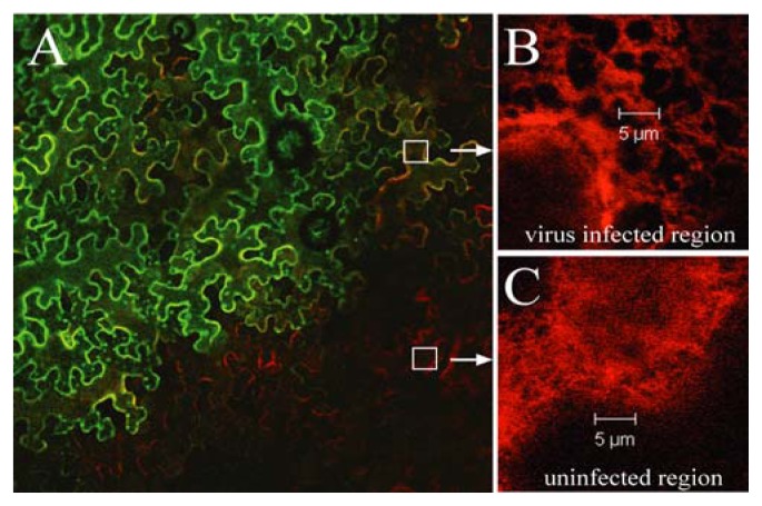
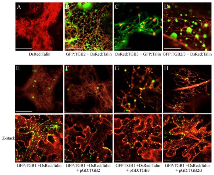
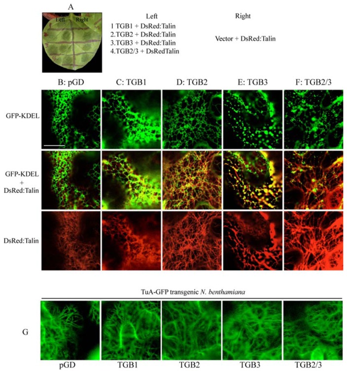
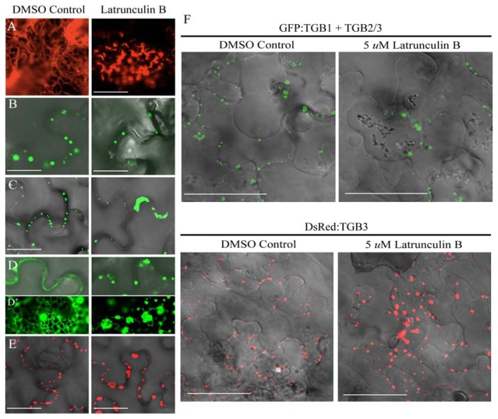
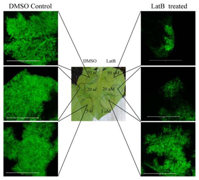
Barley stripe mosaic virus (BSMV) induces massive actin filament thickening at the infection front of infected Nicotiana benthamiana leaves. To determine the mechanisms leading to actin remodeling, fluorescent protein fusions of the BSMV triple gene block (TGB) proteins were coexpressed in cells with the actin marker DsRed: Talin. TGB ectopic expression experiments revealed that TGB3 is a major elicitor of filament thickening, that TGB2 resulted in formation of intermediate DsRed:Talin filaments, and that TGB1 alone had no obvious effects on actin filament structure. Latrunculin B (LatB) treatments retarded BSMV cell-to-cell movement, disrupted actin filament organization, and dramatically decreased the proportion of paired TGB3 foci appearing at the cell wall (CW). BSMV infection of transgenic plants tagged with GFP-KDEL exhibited membrane proliferation and vesicle formation that were especially evident around the nucleus. Similar membrane proliferation occurred in plants expressing TGB2 and/or TGB3, and DsRed: Talin fluorescence in these plants colocalized with the ER vesicles. TGB3 also associated with the Golgi apparatus and overlapped with cortical vesicles appearing at the cell periphery. Brefeldin A treatments disrupted Golgi and also altered vesicles at the CW, but failed to interfere with TGB CW localization. Our results indicate that actin cytoskeleton interactions are important in BSMV cell-to-cell movement and for CW localization of TGB3.

Keywords: Barley stripe mosaic virus; Hordeivirus; Latrunculin B; Membrane proliferation; Triple gene block.


Figures

Fig. 1
Fig. 1
Changes in actin filament structure in BSMV-infected N. benthamiana epidermal cells. (A) N. benthamiana leaf inoculated with a BSMV derivative consisting of wild-type α and β RNAs and RNAγ containing the γb:GFP fusion protein as a marker for virus infection (green). DsRed:Talin was co-expressed in the same leaf by agroinfiltration to permit visualization of actin filaments (red). Cells were examined by confocal microscopy at 3 to 10 days after inoculation, and actin filament thickness was examined at the infection fronts of cells. (B) Thick remodeled actin cables visualized by DsRed:Talin (C) DsRed:Talin expression showing the filamentous actin network in an uninvaded region of the leaf. Note: Substantially thicker actin filaments were present only in regions at or behind the infection front of regions expressing BSMV GFP. Photographs were taken at 7 dpi.
Fig. 2
Fig. 2
TGB2 and TGB3 induction of actin filament bundling and roles in TGB1 PD targeting. The actin marker protein, DsRed:Talin, was coexpressed in N. benthamiana leaves along with TGB proteins by agroinfiltration. The following combinations are shown: (A) DsRed:Talin (B) GFP:TGB2 + DsRed:Talin (C) DsRed:TGB3 + GFP:Talin (D) GFP:TGB2/3 + DsRed:Talin (E) GFP:TGB1 + DsRed:Talin (F) GFP:TGB1, TGB2 + DsRed:Talin (G) GFP:TGB1, TGB3 + DsRed:Talin (H) GFP:TGB1, TGB2/3 + DsRed:Talin. In (E-H) the top panels represent single confocal images, and the bottom panels represent a Z-stack projection of over 32 optical slices through the cell generated using the Imaris software. Bars = 20 μm. Photographs were taken at 2 days post infiltration.
Fig. 3
Fig. 3
Vesicle appearance in BSMV infected GFP-KDEL transgenic N. benthamiana epidermal cells. (A) Uninfected GFP-KDEL N. benthamiana. (B) N. benthamiana leaf inoculated with BSMV α, β, and γ RNAs. Arrows indicate vesicle formation around nucleus as a marker for virus infection (green). Cells were examined by confocal microscopy at 7 days after inoculation. Bars = 50 μm. The magnification of the exploded bottom panel exactly repeats the boxed areas in the upper panels.
Fig. 4
Fig. 4
TGB2 and TGB3 effects on ER-derived vesicles. The actin marker protein, DsRed:Talin, was expressed with the TGB proteins by agroinfiltration into GFP-KDEL transgenic N. benthamiana (A) In order to compare the effects of the TGB proteins on ER and actin within same leaf, the leaf was divided into eight sections and infiltrated with the following combinations: (B) Agrobacterium pGD vector + DsRed:Talin (C) TGB1 + DsRed:Talin (D) TGB2 + DsRed:Talin, (E) TGB3 + DsRed:Talin, or (F) TGB2/3 + DsRed:Talin. (B–F) (First panel) green channel shows GFP-KDEL (Second panel) green and red channels show GFP-KDEL and DsRed:Talin, respectively. (Third panel) red channel shows DsRed:Talin (G) The tubulin marker protein, TuA:GFP, was expressed with the TGB proteins in TuA:GFP transgenic N. benthamiana. Bar = 20 μm. Photographs were taken at 2 days post infiltration.
Fig. 5
Fig. 5
Subcellular localization and effects of TGB3 mutants on ER structure. DsRed:TGB3 mutants were expressed by agroinfiltration into GFP-KDEL transgenic N. benthamiana. Epifluorescent images of DsRed:TGB3 in unplasmolyzed cells (Left) and plasmolyzed cells (Right) were taken at 2 dpi. (A) wt TGB3 (B) TGB3(H5L, C11Y), (C) TGB3(H5L, C9Y, C11Y), (D) TGB3(Q90Y, D91R, L92Y, D93R), (E) TGB3(P105R, I108R), (F) TGB3(Q115Y, P118R, Q120R), (G) TGB3(1–150). Bar = 50 μm.
Fig. 6
Fig. 6
Cytoskeleton disruption by Latrunculin B and TGB localization. The following combinations of proteins were expressed in N. benthamiana epidermal cells by agroinfiltration: (A) DsRed:Talin, (B) GFP:TGB1 (C) GFP:TGB1 + TGB2/3 (D) GFP:TGB2 (E) DsRed:TGB3. The left panels show the localization of the proteins in the absence of Lat B, and the right panels illustrate localization of the proteins after LatB treatment. Bar = 20 μm. The insert panels (D) highlight the network occurring with TGB2. (F) Plasmolysis after LatB treatment. GFP:TGB1 + TGB2/3 and DsRed:TGB3 were expressed in the presence of DMSO (Control), or 5 μM LatB. Both treatments were visualized by confocal microscopy after plasmolysis induced by infiltration with 700 mM sucrose. Bar = 50 μm.
Fig. 7
Fig. 7
Latrunculin B effects on BSMV movement in N. benthamiana. Leaves were divided into six regions and the left side of the leaf was treated with DMSO dilutions and the right side of the leaves were infiltrated with three different concentrations of LatB (5 μm, 20 μM, or 50 μM) in DMSO. After LatB and DMSO treatments, a BSMV derivative consisting of wild-type α and β RNAs and an RNAg containing the Yb:GFP BSMV was inoculated. GFP visualization of BSMV movement was observed by confocal microscopy at 6 dpi.
Fig. 8
Fig. 8
Brefeldin A disruption of Golgi and effects on DsRed: TGB3 localization. The DsRed fusions to wtTGB3 derivatives were coexpressed with the ST:GFP Golgi marker protein in N. benthamiana epidermal cells. Cells were infiltrated with DMSO in buffer, or were treated with BFA to disrupt Golgi. Labels at the top of the columns designate whether or not cells were plasmolyzed. (A) ST-GFP, (B) DsRed:TGB3 plus ST-GFP, Bar = 20 μm. (C) Comparison of DsRed:TGB3 expression in LatB or (D) BFA treated wild type N. benthamiana after plasmolysis. Bar = 50 μm. Fluorescent images were captured at 2–3 dpi and overlayed onto DIC images.

References

    1. Amari K, Lerich A, Schmitt-Keichinger C, Dolja VV, Ritzenthaler C. Tubule-guided cell-to-cell movement of a plant virus requires class XI myosin motors. PLoS Pathol. 2011:e1002327. - PMC - PubMed
    1. Baluska F, Jasik J, Edelmann HG, Salajova T, Volkmann D. Latrunculin B-induced plant dwarfism: Plant cell elongation is F-actin-dependent. Dev Biol. 2001;231:113–124. - PubMed
    1. Boevink P, Oparka K, Santa Cruz S, Martin B, Betteridge A, Hawes C. Stacks on tracks: the plant Golgi apparatus traffics on an actin/ER network. Plant J. 1998;15:441–447. - PubMed
    1. Bragg JN, Lawrence DM, Jackson AO. The N-terminal 85 amino acids of the barley stripe mosaic virus b pathogenesis protein contain three zinc-binding motifs. J Virol. 2004;78:7379–7391. - PMC - PubMed
    1. Cowan GH, Lioliopoulou F, Ziegler A, Torrance L. Subcellular localisation, protein interactions, and RNA binding of potato mop-top virus triple gene block proteins. Virology. 2002;298:106–115. - PubMed