

Extranodal natural killer/T-cell lymphoma, nasal type, involving the skin, misdiagnosed as nasosinusitis and a fungal infection: A case report and literature review
- PMID: 25289105
- PMCID: PMC4186554
- DOI: 10.3892/ol.2014.2509
Extranodal natural killer/T-cell lymphoma, nasal type, involving the skin, misdiagnosed as nasosinusitis and a fungal infection: A case report and literature review
Abstract
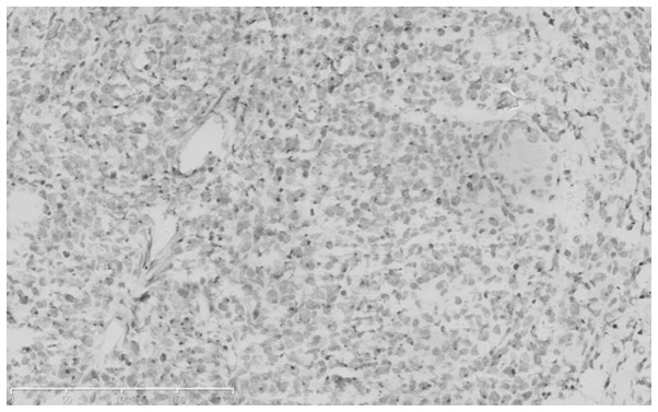
The present study reports a case of extranodal natural killer (NK)/T-cell lymphoma, nasal type, involving the skin. The clinical manifestations, pathological characteristics, treatment and prognosis of the case were analyzed to improve the clinical diagnosis and treatment for this disease. The patient was a 56-year-old male, presenting with dark red nodules and plaques that had been visible on the nose for half a year. Based on the skin lesions and histopathological and immunohistochemical examination results, the patient was diagnosed with extranodal NK/T-cell lymphoma, nasal type. This disease has unique histopathological and immunohistochemical features and a high malignancy. The condition tends to be misdiagnosed and has a poor prognosis, but seldom involves the skin. In the present case, only radiotherapy was performed, with no relapse occurring within 6 months.
Keywords: lymphoma; nasal type; natural killer/T cell; skin perforation.
Figures


Similar articles
-
Blastic natural killer cell and extranodal natural killer cell-like T-cell lymphoma presenting in the skin: report of six cases from the UK.Br J Dermatol. 2003 Mar;148(3):507-15. doi: 10.1046/j.1365-2133.2003.05227.x. Br J Dermatol. 2003. PMID: 12653743
-
Lymphomatoidgastropathy mimicking extranodal NK/T cell lymphoma, nasal type: a case report.World J Gastroenterol. 2012 May 7;18(17):2140-4. doi: 10.3748/wjg.v18.i17.2140. World J Gastroenterol. 2012. PMID: 22563204 Free PMC article.
-
Extranodal natural killer/T-cell lymphoma, nasal type: a Spanish multicentric retrospective survey.Eur J Dermatol. 2018 Feb 1;28(1):64-70. doi: 10.1684/ejd.2017.3205. Eur J Dermatol. 2018. PMID: 29400283
-
Extranodal nasal-type natural killer/T-cell lymphoma with penile involvement: a case report and review of the literature.BMC Urol. 2017 Sep 6;17(1):77. doi: 10.1186/s12894-017-0273-8. BMC Urol. 2017. PMID: 28874193 Free PMC article. Review.
-
[Extranodal T/NK-cell lymphoma, nasal type: a case report and review of the literature].An Med Interna. 2005 Dec;22(12):597-600. doi: 10.4321/s0212-71992005001200010. An Med Interna. 2005. PMID: 16454602 Review. Spanish.
Cited by
-
Extranodal Natural Killer/T-cell Lymphoma Isolated to the Leg: A Case Report.Cureus. 2023 Jun 5;15(6):e40011. doi: 10.7759/cureus.40011. eCollection 2023 Jun. Cureus. 2023. PMID: 37425545 Free PMC article.
-
Annular Erythematous Patches as the Presenting Sign of Extranodal Natural Killer/T-Cell Lymphoma.Turk J Haematol. 2016 Dec 1;33(4):360-361. doi: 10.4274/tjh.2016.0071. Epub 2016 Jul 15. Turk J Haematol. 2016. PMID: 27476759 Free PMC article. No abstract available.
-
Extranodal NK/T Cell Lymphoma with Destruction of the Uvulae: A Case Report.Iran J Otorhinolaryngol. 2017 Mar;29(91):101-108. Iran J Otorhinolaryngol. 2017. PMID: 28393058 Free PMC article.
References
-
- Swerdlow SH. WHO classification of tumours of haematopoietic and lymphoid tissues. 4th edition. Vol. 2. World Health Organization; 2008.
-
- Zhao B. China clinical skin disease science. Jiangsu Science and Technology Press. 2009;1661:1679–1681.
-
- Li S, Feng X, Li T, et al. Extranodal NK/T-cell lymphoma, nasal type: a report of 73 cases at MD Anderson Cancer Center. Am J Surg Pathol. 2013;37:14–23. - PubMed
-
- Li S, Feng X, Li T, et al. Extranodal NK/T-cell lymphoma, nasal type: a report of 73 cases at MD Anderson Cancer Center. Am J Surg Pathol. 2013;37:14–23. - PubMed
-
- Lister TA, Crowther D, Sutcliffe SB, et al. Report of a committee convened to discuss the evaluation and staging of patients with Hodgkin’s disease: Cotswolds meeting. J Clin Oncol. 1989;7:1630–1636. - PubMed
LinkOut - more resources
Full Text Sources
Other Literature Sources