. 2013 Jun 7;1(2):1-9.
doi: 10.1097/GOX.0b013e318293dc69. eCollection 2013 May.

Double-eyelid surgery using septoaponeurosis junctional thickening results in dynamic fold in asians

Affiliations

Double-eyelid surgery using septoaponeurosis junctional thickening results in dynamic fold in asians

Hong Seok Kim et al. Plast Reconstr Surg Glob Open. .

Abstract

Background: To avoid a static double-eyelid fold characterized by nonmobile overdepression of the fold, we propose a new surgical approach of using septoaponeurosis junctional thickening (SAJT) to create a dynamic fold.

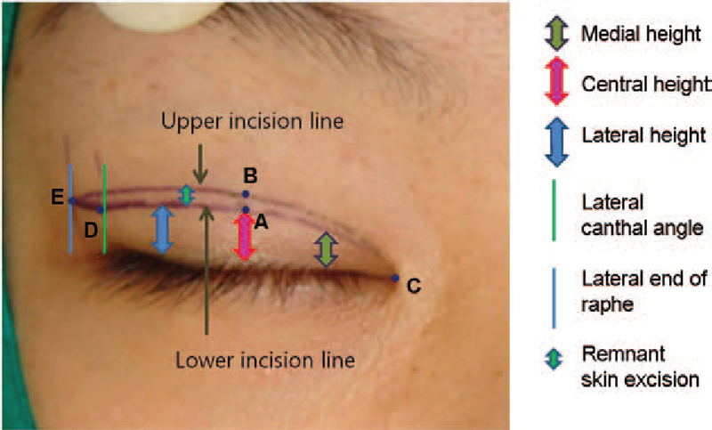
Methods: Six hundred eighty patients underwent double-eyelid surgery using the SAJT fixation technique. The orbital septum was exposed and transversely opened superior to the incision margin. The lower septal stump was trimmed to expose the SAJT. The dermis and orbicularis oculi muscle of the lower flap of the upper eyelid were attached to the SAJT. Patients were followed for 2-8 years (mean, 3.6 y). Anatomic study with 28 upper eyelids from 28 Korean adult cadavers was performed to confirm the histological structure of the SAJT.

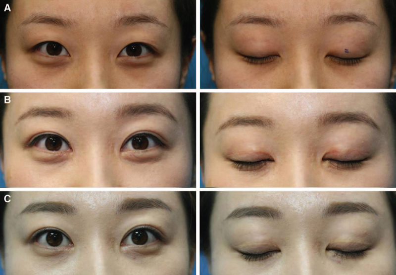
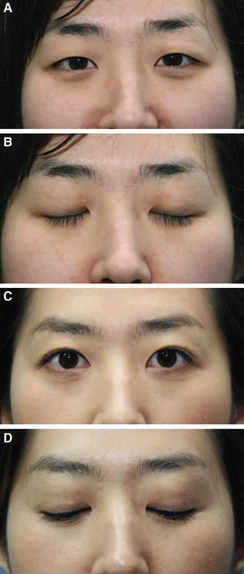
Results: This technique created a dynamic fold. When the eyes were open, the fold depth was moderate. When the eyes were closed, the fold site was smooth and not depressed. The surgery had a 95% patient satisfaction rate (365 responded as satisfied and 236 responded as very satisfied). Postoperative complications included partial or complete loss of the double-eyelid line in 14 and 4 cases, respectively, hypertrophic scar formation in 7 cases, and asymmetric fold in 8 cases.

Conclusions: The authors introduce a double-eyelid surgery technique using the SAJT. This SAJT fixation technique creates a dynamic double-eyelid fold. Our study showed a high patient satisfaction rate and that the resulting fold mimics the movement of the congenital supratarsal fold in Asians.


Figures

Fig. 1.
Fig. 1.
Photographs of a 27-y-old woman exhibiting a static fold after she underwent double-eyelid surgery using the tarsal fixation technique. A static fold is characterized by deep fold formation (A), a visibly depressed line along the incision with eyes closed, and an immobile line (B).
Video Graphic 1.
Video Graphic 1.
See video, Supplemental Digital Content 1, which shows the front and side views of a 29-year-old woman exhibiting a static fold after she underwent double eyelid surgery using the tarsal fixation technique as she opens and closes her eyes, http://links.lww.com/PRSGO/A0.
Fig. 2.
Fig. 2.
A 24-y-old woman who underwent the incisional double-eyelid surgery technique (A). Two years postoperatively, the patient is exhibiting a dynamic fold, which is characterized by moderate depth of the fold with eyes open (B) and a minimally depressed scar along the incision line with eyes closed (C) with a mobile fold line.
Video Graphic 2.
Video Graphic 2.
See video, Supplemental Digital Content 2, which shows the front and quarter views of the same patient exhibiting a dynamic fold after she received double eyelid surgery using septoaponeurosis junctional thickening fixation as she opens and closes her eyes, http://links.lww.com/PRSGO/A1.
Fig. 3.
Fig. 3.
Histology showing the SAJT, the inner layer (red arrow) and outer layer (blue arrow) of the septum, levator aponeurosis (LA) and the tarsus (A). The SAJT lies in between the outer layers of the orbital septum and the LA (B).
Fig. 4.
Fig. 4.
Preoperative designing of the lower and upper incision lines: Central height (A), extent of skin excision (B), medial canthus (C), lateral height (D), and lateral raphe (E).
Fig. 5.
Fig. 5.
(A) The septum was transversely incised at 7 mm above the skin incision site using scissors from lateral to medial direction. (B) Complete incision of the septum is required to prevent hindrance of LA movement while uncovering the SAJT.
Fig. 6.
Fig. 6.
(A) The lower stump of the septum was trimmed before suture fixation. (B) The remnant of the septum was pulled and examined to determine whether the SAJT was adequately trimmed.
Fig. 7.
Fig. 7.
Diagram showing the suture fixation of the dermis and SAJT. (A) The SAJT defines the thickened septal tissue between the inner (red arrow) and outer layers (blue arrow) of the orbital septum immediately cephalad to the tarsus and anterior to the LA. (B) Once the SAJT was uncovered and trimmed, the dermis of the lower flap was fixated to the SAJT at 6–8 sites. (C) The skin closure was performed with 8-0 black nylon in a continuous running fashion.
Fig. 8.
Fig. 8.
Preoperative view of a 28-y-old woman with redundant and puffy upper eyelids with eyes open (left column) and with eyes closed (right column) (A). The patient underwent incisional double-eyelid surgery using SAJT fixation and epicanthoplasty. One-month (B) and 4-y (C) postoperative views with eyes open (left column) and eyes closed (right column).
Fig. 9.
Fig. 9.
Preoperative view of a 22-y-old woman with redundant and puffy upper eyelids with eyes open (A) and with eyes closed (B). Three-year postoperative view with eyes open (C) and with eyes closed (D).

References

    1. Chen W PD. Asian Blepharoplasty and the Eyelid Crease. 2nd ed. New York, NY: Elsevier; 2006.
    1. McCurdy JA. Cosmetic Surgery of the Asian Face. New York, NY: Thieme Medical Publishers; 1990.
    1. Kikkawa DO, Kim JW. Asian blepharoplasty. Int Ophthalmol Clin. 1997;37:193–204. - PubMed
    1. Chen W PD. In: In: Cosmetic Oculoplastic Surgery. Putterman AM, editor. Philadelphia, PA: WB Saunders; 1999. pp. 101–111.
    1. Jeong S, Lemke BN, Dortzbach RK, et al. The Asian upper eyelid: an anatomical study with comparison to the Caucasian eyelid. Arch Ophthalmol. 1999;117:907–912. - PubMed

LinkOut - more resources