. 2013 Oct 7;1(6):e41.
doi: 10.1097/GOX.0b013e3182a7f222. eCollection 2013 Sep.

Histological and Ultrastructural Effects of Ultrasound-induced Cavitation on Human Skin Adipose Tissue

Affiliations

Histological and Ultrastructural Effects of Ultrasound-induced Cavitation on Human Skin Adipose Tissue

Daniele Bani et al. Plast Reconstr Surg Glob Open. .

Abstract

Background: In aesthetic medicine, the most promising techniques for noninvasive body sculpturing purposes are based on ultrasound-induced fat cavitation. Liporeductive ultrasound devices afford clinically relevant subcutaneous fat pad reduction without significant adverse reactions. This study aims at evaluating the histological and ultrastructural changes induced by ultrasound cavitation on the different cell components of human skin.

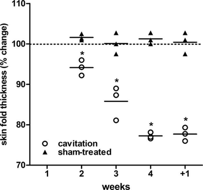
Methods: Control and ultrasound-treated ex vivo abdominal full-thickness skin samples and skin biopsies from patients pretreated with or without ultrasound cavitation were studied histologically, morphometrically, and ultrastructurally to evaluate possible changes in adipocyte size and morphology. Adipocyte apoptosis and triglyceride release were also assayed. Clinical evaluation of the effects of 4 weekly ultrasound vs sham treatments was performed by plicometry.

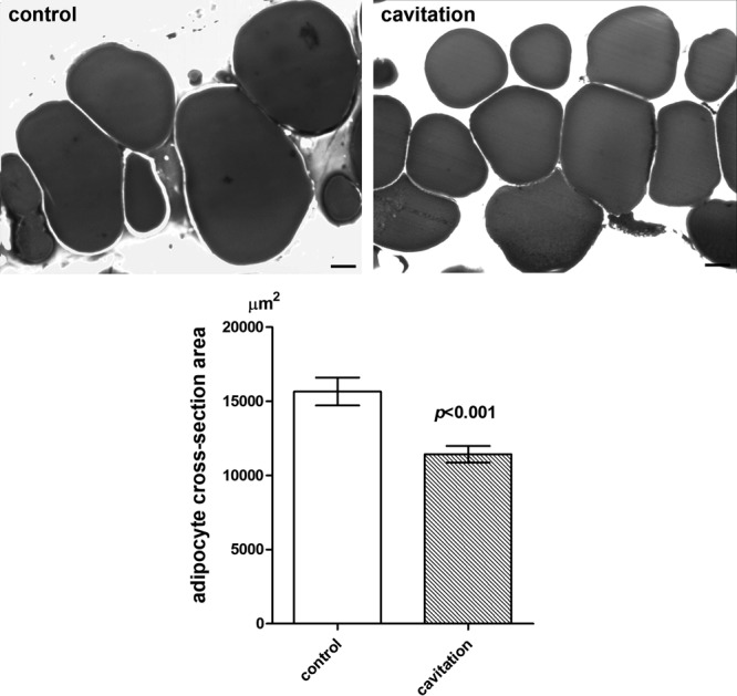
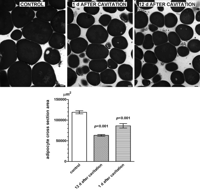
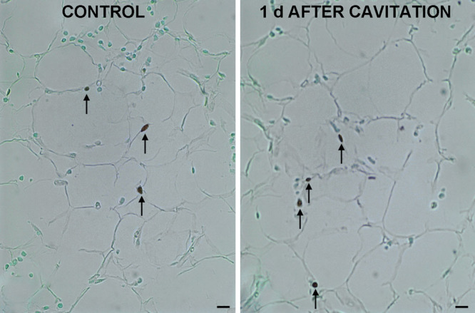
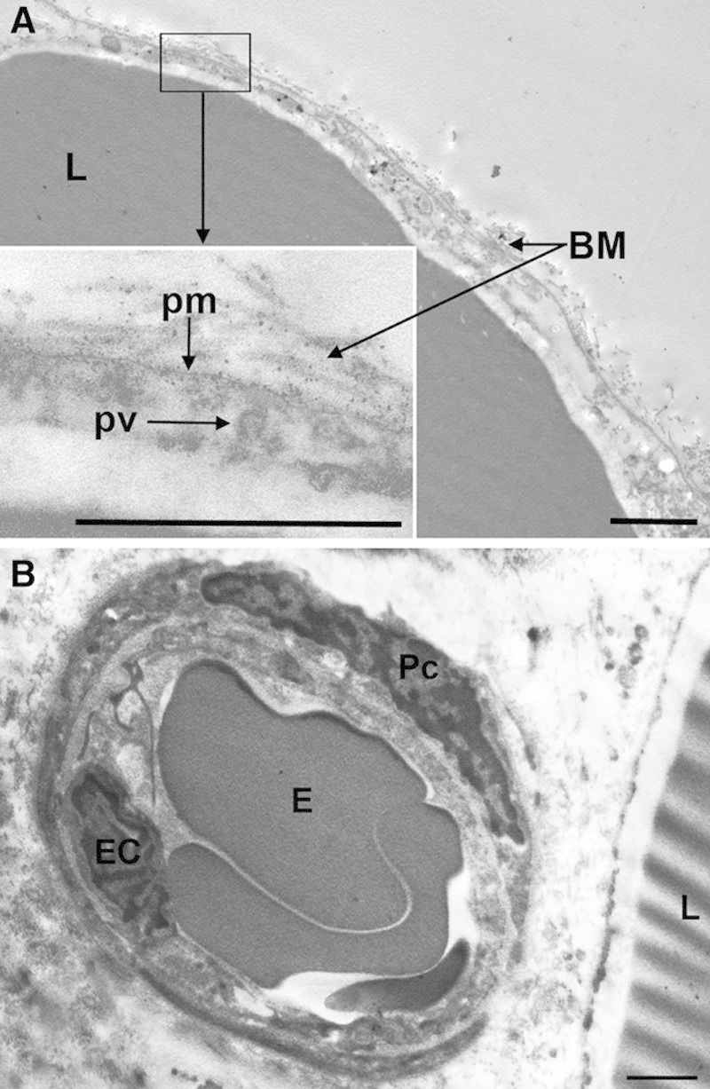
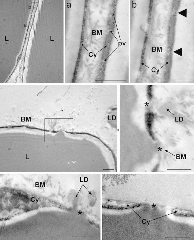
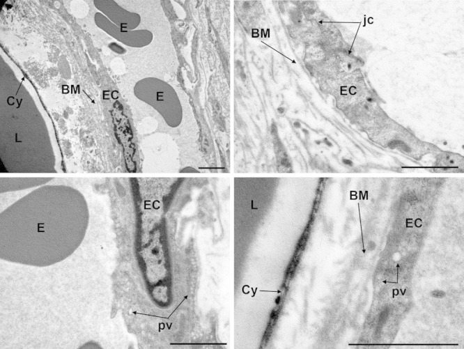
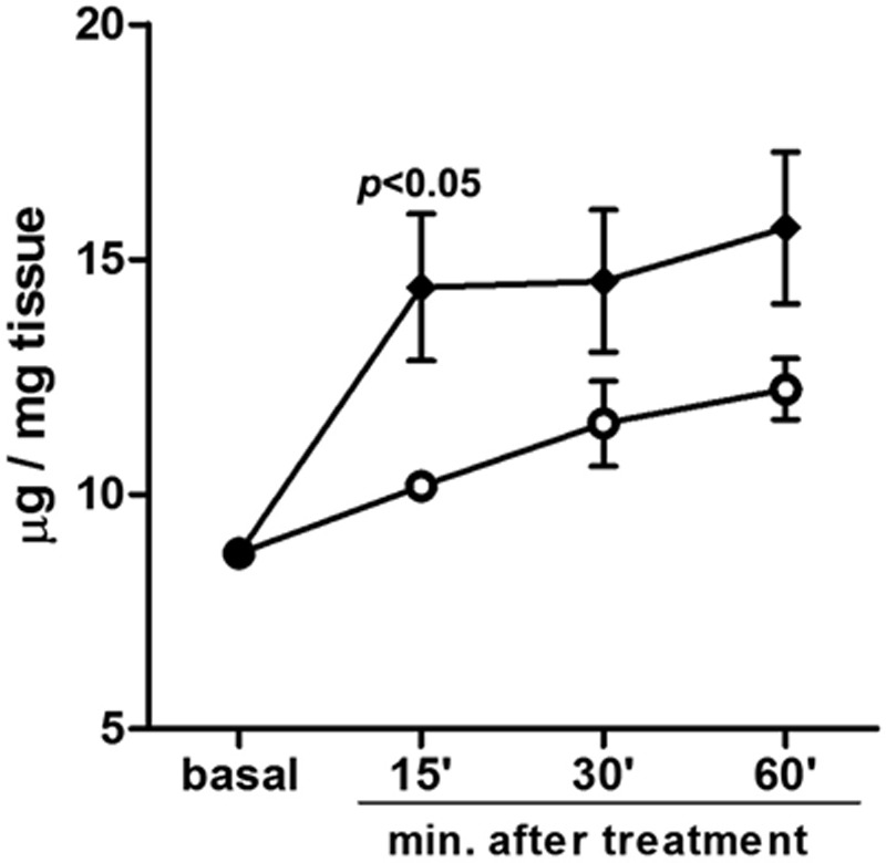
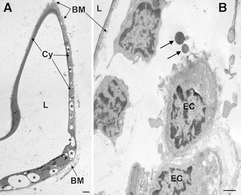
Results: Compared with the sham-treated control samples, ultrasound cavitation induced a statistically significant reduction in the size of the adipocytes (P < 0.001), the appearance of micropores and triglyceride leakage and release in the conditioned medium (P < 0.05 at 15 min), or adipose tissue interstitium, without appreciable changes in microvascular, stromal, and epidermal components and in the number of apoptotic adipocytes. Clinically, the ultrasound treatment caused a significant reduction of abdominal fat.

Conclusions: This study further strengthens the current notion that noninvasive transcutaneous ultrasound cavitation is a promising and safe technology for localized reduction of fat and provides experimental evidence for its specific mechanism of action on the adipocytes.


Figures

Fig. 1.
Fig. 1.
Histological and morphometrical findings of subcutaneous adipocytes from ex vivo skin explants. Ultrasound cavitation causes a statistically significant reduction of mean cross-section surface area of lipid vacuoles, related to adipocyte overall volume (Student’s t test, n = 3 biopsies per experimental group). OsO4 fixation/staining and toluidine blue counterstaining. Bars = 10 µm.
Fig. 2.
Fig. 2.
Histological and morphometrical findings of subcutaneous adipocytes from control or ultrasound-pretreated abdominal skin biopsies taken at the noted times after the treatment. Ultrasound cavitation causes a statistically significant reduction of mean cross-section surface area of lipid vacuoles, related to adipocyte overall volume (Student’s t test, n = 10 microscopical fields per biopsy). No differences were observed between the control samples from the 2 patients. OsO4 fixation/staining and toluidine blue counterstaining. Bars = 10 µm.
Fig. 3.
Fig. 3.
TUNEL assay performed to detect apoptotic adipocytes in subcutaneous biopsies from sham- or ultrasound-pretreated abdominal skin. Arrows point at apoptotic nuclei. No differences were observed or measured between the 2 groups. Bars = 10 µm.
Fig. 4.
Fig. 4.
Representative transmission electron microscopy images of adipose tissue from sham-treated ex vivo skin explants. A, Adipocytes show normal features. B, Control adipose tissue interstitium also shows normal morphology. BM, basement membrane; E, erythrocyte; EC, endothelial cell; L, lipid vacuoles; Pc, pericyte; pm, plasma membrane; pv, pinocytosis vesicle. Bars = 1 µm.
Fig. 5.
Fig. 5.
Representative transmission electron microscopy images of adipose tissue from ultrasound-treated ex vivo skin explants. Upper panels: Two adjacent adipocytes are shown. BM, basement membranes; Cy, cytoplasm; L, lipid vacuoles; pv, pinocytosis vesicles. The arrowheads in the inset (b) point at 2 areas, featuring microvesicular clusters, in which the cytoplasm appears to be about to rupture. Centre and lower panels: Abnormal adipocytes undergoing lipid release. L, lipid vacuoles; LD, lipid droplets in the extracellular matrix. The asterisks indicate focal cytoplasmic ruptures allowing leakage of triglyceride droplets in the interstitium. Bars = 1 µm.
Fig. 6.
Fig. 6.
Representative transmission electron microscopy images of adipose tissue from ultrasound-treated ex vivo skin explants. Endothelial cells (EC) of blood microvessels show normal features. BM, basement membrane; Cy, adipocyte cytoplasm; E, erythrocytes; jc, junctional complex; L, lipid vacuoles; pv, pinocytosis vesicles. Bars = 1 µm.
Fig. 7.
Fig. 7.
Assay of triglycerides released in the conditioned medium of ex vivo skin explants. A time-related increase is observed in both the sham- and ultrasound-treated specimens. Cavitation induced a higher release of triglycerides as compared with the controls, peaking at 15 min.
Fig. 8.
Fig. 8.
Representative transmission electron microscopy images of adipose tissue from ultrasound-pretreated (1 d) abdominal skin areas. A, An adipocyte showing irregular, winding profiles and multiple lipid droplets (some of which are labeled by asterisks) clustered in the cytoplasmic rim. B, Adipose tissue interstitium showing normal blood microvessels and free lipid droplets (arrows) in their proximity. BM, basement membrane; Cy, adipocyte cytoplasm; EC, endothelial cells; L, lipid vacuoles. Bars = 1 µm.
Fig. 9.
Fig. 9.
Percent changes of the thickness of sham- and ultrasound-treated abdominal skin folds measured by plicometry upon 4 weekly treatments and 1 wk after the last treatment (+1). A time-related, statistically significant decrease is observed in the ultrasound-treated skin areas as compared with the sham-treated controls. Two-way analysis of variance and Bonferroni’s posttest: n = 3, *P < 0.001.

References

    1. Housman TS, Lawrence N, Mellen BG, et al. The safety of liposuction: results of a national survey. Dermatol Surg. 2002;28:971–978. - PubMed
    1. Coleman KM, Coleman WP, III, Benchetrit A. Non-invasive, external ultrasonic lipolysis. Semin Cutan Med Surg. 2009;28:263–267. - PubMed
    1. Neira R, Arroyave J, Ramirez H, et al. Fat liquefaction: effect of low-level laser energy on adipose tissue. Plast Reconstr Surg. 2002;110:912–922; discussion 923. - PubMed
    1. Jackson RF, Dedo DD, Roche GC, et al. Low-level laser therapy as a non-invasive approach for body contouring: a randomized controlled study. Lasers Surg Med. 2009;41:799–809. - PubMed
    1. Manuskiatti W, Wachirakaphan C, Lektrakul N, et al. Circumference reduction and cellulite treatment with a Tri-Polar radiofrequency device: a pilot study. J Eur Acad Dermatol Venereol. 2009;23:820–827. - PubMed